ARG63129
anti-FGR antibody
anti-FGR antibody for Western blot and Human
Cancer antibody; Signaling Transduction antibody
Overview
| Product Description | Goat Polyclonal antibody recognizes FGR |
|---|---|
| Tested Reactivity | Hu |
| Predict Reactivity | Ms, Rat, Dog |
| Tested Application | WB |
| Specificity | This antibody is expected to recognise all three reported isoforms (NP_001036194.1; NP_001036212.1; NP_005239.1). |
| Host | Goat |
| Clonality | Polyclonal |
| Isotype | IgG |
| Target Name | FGR |
| Antigen Species | Human |
| Immunogen | C-TSAEPQYQPGDQT |
| Conjugation | Un-conjugated |
| Alternate Names | p58-Fgr; v-fgr; Tyrosine-protein kinase Fgr; p55-Fgr; p58c-Fgr; SRC2; Proto-oncogene c-Fgr; p55c-fgr; p58c-fgr; Gardner-Rasheed feline sarcoma viral; c-fgr; c-src2; EC 2.7.10.2 |
Application Instructions
| Application Suggestion |
|
||||
|---|---|---|---|---|---|
| Application Note | WB: Recommend incubate at RT for 1h. * The dilutions indicate recommended starting dilutions and the optimal dilutions or concentrations should be determined by the scientist. |
Properties
| Form | Liquid |
|---|---|
| Purification | Purified from goat serum by antigen affinity chromatography. |
| Buffer | Tris saline (pH 7.3), 0.02% Sodium azide and 0.5% BSA. |
| Preservative | 0.02% Sodium azide |
| Stabilizer | 0.5% BSA |
| Concentration | 0.5 mg/ml |
| Storage Instruction | For continuous use, store undiluted antibody at 2-8°C for up to a week. For long-term storage, aliquot and store at -20°C or below. Storage in frost free freezers is not recommended. Avoid repeated freeze/thaw cycles. Suggest spin the vial prior to opening. The antibody solution should be gently mixed before use. |
| Note | For laboratory research only, not for drug, diagnostic or other use. |
Bioinformation
| Database Links | |
|---|---|
| Background | This gene is a member of the Src family of protein tyrosine kinases (PTKs). The encoded protein contains N-terminal sites for myristylation and palmitylation, a PTK domain, and SH2 and SH3 domains which are involved in mediating protein-protein interactions with phosphotyrosine-containing and proline-rich motifs, respectively. The protein localizes to plasma membrane ruffles, and functions as a negative regulator of cell migration and adhesion triggered by the beta-2 integrin signal transduction pathway. Infection with Epstein-Barr virus results in the overexpression of this gene. Multiple alternatively spliced variants, encoding the same protein, have been identified. [provided by RefSeq, Jul 2008] |
| Research Area | Cancer antibody; Signaling Transduction antibody |
| Calculated MW | 59 kDa |
| PTM | Ubiquitinated. Becomes ubiquitinated in response to ITGB2 signaling; this does not lead to degradation. Phosphorylated. Autophosphorylated on tyrosine residues. Becomes phosphorylated in response to FCGR2A and/or FCGR2B engagement, cell adhesion and signaling by ITGB2. Prior phosphorylation at Tyr-523 by SRC inhibits ulterior autophosphorylation at Tyr-412. |
Images (2) Click the Picture to Zoom In
-
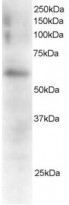
ARG63129 anti-FGR antibody WB image
Western Blot: Mouse Spleen extracts (RIPA buffer, 35 µg total protein per lane) stained with ARG63129 anti-FGR antibody at 0.5 µg/ml dilution.
-
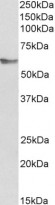
ARG63129 anti-FGR antibody WB image
Western blot: 35 µg of Human peripheral blood lymphocyte lysates (in RIPA buffer) stained with ARG63129 anti-FGR antibody at 0.3 µg/ml dilution and incubated at RT for 1 hour.


