ARG55365
anti-FGR antibody
anti-FGR antibody for ICC/IF,IHC-Formalin-fixed paraffin-embedded sections,Western blot and Human
Cancer antibody; Signaling Transduction antibody
Overview
| Product Description | Rabbit Polyclonal antibody recognizes FGR |
|---|---|
| Tested Reactivity | Hu |
| Tested Application | ICC/IF, IHC-P, WB |
| Host | Rabbit |
| Clonality | Polyclonal |
| Isotype | IgG |
| Target Name | FGR |
| Antigen Species | Human |
| Immunogen | KLH-conjugated synthetic peptide corresponding to aa. 3-33 (N-terminus) of Human FGR. |
| Conjugation | Un-conjugated |
| Alternate Names | p58-Fgr; v-fgr; Tyrosine-protein kinase Fgr; p55-Fgr; p58c-Fgr; SRC2; Proto-oncogene c-Fgr; p55c-fgr; p58c-fgr; Gardner-Rasheed feline sarcoma viral; c-fgr; c-src2; EC 2.7.10.2 |
Application Instructions
| Application Suggestion |
|
||||||||
|---|---|---|---|---|---|---|---|---|---|
| Application Note | * The dilutions indicate recommended starting dilutions and the optimal dilutions or concentrations should be determined by the scientist. | ||||||||
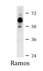
| Positive Control | Ramos |
Properties
| Form | Liquid |
|---|---|
| Purification | Purification with Protein G. |
| Buffer | PBS and 0.09% (W/V) Sodium azide |
| Preservative | 0.09% (W/V) Sodium azide |
| Storage Instruction | For continuous use, store undiluted antibody at 2-8°C for up to a week. For long-term storage, aliquot and store at -20°C or below. Storage in frost free freezers is not recommended. Avoid repeated freeze/thaw cycles. Suggest spin the vial prior to opening. The antibody solution should be gently mixed before use. |
| Note | For laboratory research only, not for drug, diagnostic or other use. |
Bioinformation
| Database Links | |
|---|---|
| Gene Symbol | FGR |
| Gene Full Name | FGR proto-oncogene, Src family tyrosine kinase |
| Background | This gene is a member of the Src family of protein tyrosine kinases (PTKs). The encoded protein contains N-terminal sites for myristylation and palmitylation, a PTK domain, and SH2 and SH3 domains which are involved in mediating protein-protein interactions with phosphotyrosine-containing and proline-rich motifs, respectively. The protein localizes to plasma membrane ruffles, and functions as a negative regulator of cell migration and adhesion triggered by the beta-2 integrin signal transduction pathway. Infection with Epstein-Barr virus results in the overexpression of this gene. Multiple alternatively spliced variants, encoding the same protein, have been identified. [provided by RefSeq, Jul 2008] |
| Function | Non-receptor tyrosine-protein kinase that transmits signals from cell surface receptors devoid of kinase activity and contributes to the regulation of immune responses, including neutrophil, monocyte, macrophage and mast cell functions, cytoskeleton remodeling in response to extracellular stimuli, phagocytosis, cell adhesion and migration. Promotes mast cell degranulation, release of inflammatory cytokines and IgE-mediated anaphylaxis. Acts downstream of receptors that bind the Fc region of immunoglobulins, such as MS4A2/FCER1B, FCGR2A and/or FCGR2B. Acts downstream of ITGB1 and ITGB2, and regulates actin cytoskeleton reorganization, cell spreading and adhesion. Depending on the context, activates or inhibits cellular responses. Functions as negative regulator of ITGB2 signaling, phagocytosis and SYK activity in monocytes. Required for normal ITGB1 and ITGB2 signaling, normal cell spreading and adhesion in neutrophils and macrophages. Functions as positive regulator of cell migration and regulates cytoskeleton reorganization via RAC1 activation. Phosphorylates SYK (in vitro) and promotes SYK-dependent activation of AKT1 and MAP kinase signaling. Phosphorylates PLD2 in antigen-stimulated mast cells, leading to PLD2 activation and the production of the signaling molecules lysophosphatidic acid and diacylglycerol. Promotes activation of PIK3R1. Phosphorylates FASLG, and thereby regulates its ubiquitination and subsequent internalization. Phosphorylates ABL1. Promotes phosphorylation of CBL, CTTN, PIK3R1, PTK2/FAK1, PTK2B/PYK2 and VAV2. Phosphorylates HCLS1 that has already been phosphorylated by SYK, but not unphosphorylated HCLS1. [UniProt] |
| Cellular Localization | Cell membrane; Lipid-anchor; Cytoplasmic side. Cell membrane; Peripheral membrane protein; Cytoplasmic side. Cell projection, ruffle membrane. Cytoplasm, cytosol. Cytoplasm, cytoskeleton Mitochondrion inner membrane. Mitochondrion intermembrane space. Note=Detected in mitochondrial intermembrane space and at inner membranes (By similarity) Colocalizes with actin fibers at membrane ruffles. Detected at plasma membrane lipid rafts. |
| Research Area | Cancer antibody; Signaling Transduction antibody |
| Calculated MW | 59 kDa |
| PTM | Ubiquitinated. Becomes ubiquitinated in response to ITGB2 signaling; this does not lead to degradation. Phosphorylated. Autophosphorylated on tyrosine residues. Becomes phosphorylated in response to FCGR2A and/or FCGR2B engagement, cell adhesion and signaling by ITGB2. Prior phosphorylation at Tyr-523 by SRC inhibits ulterior autophosphorylation at Tyr-412. |
Images (3) Click the Picture to Zoom In
-
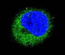
ARG55365 anti-FGR antibody ICC/IF image
Immunofluorescence: MDA-MB-231 cells stained with ARG55365 anti-FGR antibody (green). DAPI (blue) for nuclear staining.
-
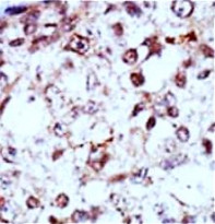
ARG55365 anti-FGR antibody IHC-P image
Immunohistochemistry: Formalin-fixed and paraffin-embedded Human cancer tissue stained with ARG55365 anti-FGR antibody.
-

ARG55365 anti-FGR antibody WB image
Western blot: Ramos cell lysate stained with ARG55365 anti-FGR antibody.