ARG41536
anti-FGFR3 antibody
anti-FGFR3 antibody for Western blot and Human,Mouse,Rat
Overview
| Product Description | Rabbit Polyclonal antibody recognizes FGFR3 |
|---|---|
| Tested Reactivity | Hu, Ms, Rat |
| Tested Application | WB |
| Host | Rabbit |
| Clonality | Polyclonal |
| Isotype | IgG |
| Target Name | FGFR3 |
| Antigen Species | Human |
| Immunogen | Synthetic peptide of Human FGFR3. |
| Conjugation | Un-conjugated |
| Alternate Names | CEK2; CD antigen CD333; FGFR-3; ACH; JTK4; Fibroblast growth factor receptor 3; CD333; EC 2.7.10.1; HSFGFR3EX |
Application Instructions
| Application Suggestion |
|
||||
|---|---|---|---|---|---|
| Application Note | * The dilutions indicate recommended starting dilutions and the optimal dilutions or concentrations should be determined by the scientist. | ||||
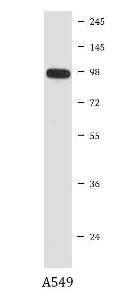
| Positive Control | A549 | ||||
| Observed Size | ~ 98 kDa |
Properties
| Form | Liquid |
|---|---|
| Purification | Affinity purified. |
| Buffer | PBS (pH 7.4), 150 mM NaCl, 0.02% Sodium azide and 50% Glycerol. |
| Preservative | 0.02% Sodium azide |
| Stabilizer | 50% Glycerol |
| Storage Instruction | For continuous use, store undiluted antibody at 2-8°C for up to a week. For long-term storage, aliquot and store at -20°C. Storage in frost free freezers is not recommended. Avoid repeated freeze/thaw cycles. Suggest spin the vial prior to opening. The antibody solution should be gently mixed before use. |
| Note | For laboratory research only, not for drug, diagnostic or other use. |
Bioinformation
| Database Links |
Swiss-port # P22607 Human Fibroblast growth factor receptor 3 |
|---|---|
| Gene Symbol | FGFR3 |
| Gene Full Name | fibroblast growth factor receptor 3 |
| Background | This gene encodes a member of the fibroblast growth factor receptor (FGFR) family, with its amino acid sequence being highly conserved between members and among divergent species. FGFR family members differ from one another in their ligand affinities and tissue distribution. A full-length representative protein would consist of an extracellular region, composed of three immunoglobulin-like domains, a single hydrophobic membrane-spanning segment and a cytoplasmic tyrosine kinase domain. The extracellular portion of the protein interacts with fibroblast growth factors, setting in motion a cascade of downstream signals, ultimately influencing mitogenesis and differentiation. This particular family member binds acidic and basic fibroblast growth hormone and plays a role in bone development and maintenance. Mutations in this gene lead to craniosynostosis and multiple types of skeletal dysplasia. Three alternatively spliced transcript variants that encode different protein isoforms have been described. [provided by RefSeq, Jul 2009] |
| Function | Tyrosine-protein kinase that acts as cell-surface receptor for fibroblast growth factors and plays an essential role in the regulation of cell proliferation, differentiation and apoptosis. Plays an essential role in the regulation of chondrocyte differentiation, proliferation and apoptosis, and is required for normal skeleton development. Regulates both osteogenesis and postnatal bone mineralization by osteoblasts. Promotes apoptosis in chondrocytes, but can also promote cancer cell proliferation. Required for normal development of the inner ear. Phosphorylates PLCG1, CBL and FRS2. Ligand binding leads to the activation of several signaling cascades. Activation of PLCG1 leads to the production of the cellular signaling molecules diacylglycerol and inositol 1,4,5-trisphosphate. Phosphorylation of FRS2 triggers recruitment of GRB2, GAB1, PIK3R1 and SOS1, and mediates activation of RAS, MAPK1/ERK2, MAPK3/ERK1 and the MAP kinase signaling pathway, as well as of the AKT1 signaling pathway. Plays a role in the regulation of vitamin D metabolism. Mutations that lead to constitutive kinase activation or impair normal FGFR3 maturation, internalization and degradation lead to aberrant signaling. Over-expressed or constitutively activated FGFR3 promotes activation of PTPN11/SHP2, STAT1, STAT5A and STAT5B. Secreted isoform 3 retains its capacity to bind FGF1 and FGF2 and hence may interfere with FGF signaling. [UniProt] |
| Cellular Localization | Isoform 1: Cell membrane; Single-pass type I membrane protein. Cytoplasmic vesicle. Endoplasmic reticulum. Note=The activated receptor is rapidly internalized and degraded. Detected in intracellular vesicles after internalization of the autophosphorylated receptor. Isoform 2: Cell membrane; Single-pass type I membrane protein. Isoform 3: Secreted. Isoform 4: Cell membrane; Single-pass type I membrane protein. [UniProt] |
| Calculated MW | 88 kDa |
| PTM | Autophosphorylated. Binding of FGF family members together with heparan sulfate proteoglycan or heparin promotes receptor dimerization and autophosphorylation on tyrosine residues. Autophosphorylation occurs in trans between the two FGFR molecules present in the dimer. Phosphorylation at Tyr-724 is essential for stimulation of cell proliferation and activation of PIK3R1, STAT1 and MAP kinase signaling. Phosphorylation at Tyr-760 is required for interaction with PIK3R1 and PLCG1. Ubiquitinated. Is rapidly ubiquitinated after ligand binding and autophosphorylation, leading to receptor internalization and degradation. Subject to both proteasomal and lysosomal degradation. N-glycosylated in the endoplasmic reticulum. The N-glycan chains undergo further maturation to an Endo H-resistant form in the Golgi apparatus. [UniProt] |
Images (1) Click the Picture to Zoom In
-
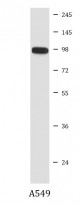
ARG41536 anti-FGFR3 antibody WB image
Western blot: A549 cell lysate stained with ARG41536 anti-FGFR3 antibody.