ARG64673
anti-FGFR2 antibody
anti-FGFR2 antibody for Western blot and Human
Cancer antibody; Cell Biology and Cellular Response antibody; Controls and Markers antibody; Developmental Biology antibody; Neuroscience antibody; Signaling Transduction antibody
Overview
| Product Description | Goat Polyclonal antibody recognizes FGFR2 |
|---|---|
| Tested Reactivity | Hu |
| Predict Reactivity | Ms, Rat, Cow, Dog, Pig |
| Tested Application | WB |
| Specificity | This antibody is expected to recognise all reported isoforms except isoform 7 (NP_001138389.1). The immunizing peptide represents part of the extracellular domain. |
| Host | Goat |
| Clonality | Polyclonal |
| Isotype | IgG |
| Target Name | FGFR2 |
| Antigen Species | Human |
| Immunogen | C-GREKEITASPDY |
| Conjugation | Un-conjugated |
| Alternate Names | CD antigen CD332; BEK; Keratinocyte growth factor receptor; K-SAM; ECT1; FGFR-2; KGFR; JWS; TK14; CFD1; BBDS; TK25; K-sam; CEK3; Fibroblast growth factor receptor 2; EC 2.7.10.1; CD332; BFR-1 |
Application Instructions
| Application Suggestion |
|
||||
|---|---|---|---|---|---|
| Application Note | WB: Recommend incubate at RT for 1h. * The dilutions indicate recommended starting dilutions and the optimal dilutions or concentrations should be determined by the scientist. |
Properties
| Form | Liquid |
|---|---|
| Purification | Purified from goat serum by antigen affinity chromatography. |
| Buffer | Tris saline (pH 7.3), 0.02% Sodium azide and 0.5% BSA. |
| Preservative | 0.02% Sodium azide |
| Stabilizer | 0.5% BSA |
| Concentration | 0.5 mg/ml |
| Storage Instruction | For continuous use, store undiluted antibody at 2-8°C for up to a week. For long-term storage, aliquot and store at -20°C or below. Storage in frost free freezers is not recommended. Avoid repeated freeze/thaw cycles. Suggest spin the vial prior to opening. The antibody solution should be gently mixed before use. |
| Note | For laboratory research only, not for drug, diagnostic or other use. |
Bioinformation
| Database Links |
Swiss-port # P21802 Human Fibroblast growth factor receptor 2 |
|---|---|
| Background | The protein encoded by this gene is a member of the fibroblast growth factor receptor family, where amino acid sequence is highly conserved between members and throughout evolution. FGFR family members differ from one another in their ligand affinities and tissue distribution. A full-length representative protein consists of an extracellular region, composed of three immunoglobulin-like domains, a single hydrophobic membrane-spanning segment and a cytoplasmic tyrosine kinase domain. The extracellular portion of the protein interacts with fibroblast growth factors, setting in motion a cascade of downstream signals, ultimately influencing mitogenesis and differentiation. This particular family member is a high-affinity receptor for acidic, basic and/or keratinocyte growth factor, depending on the isoform. Mutations in this gene are associated with Crouzon syndrome, Pfeiffer syndrome, Craniosynostosis, Apert syndrome, Jackson-Weiss syndrome, Beare-Stevenson cutis gyrata syndrome, Saethre-Chotzen syndrome, and syndromic craniosynostosis. Multiple alternatively spliced transcript variants encoding different isoforms have been noted for this gene. [provided by RefSeq, Jan 2009] |
| Research Area | Cancer antibody; Cell Biology and Cellular Response antibody; Controls and Markers antibody; Developmental Biology antibody; Neuroscience antibody; Signaling Transduction antibody |
| Calculated MW | 92 kDa |
| PTM | Autophosphorylated. Binding of FGF family members together with heparan sulfate proteoglycan or heparin promotes receptor dimerization and autophosphorylation on several tyrosine residues. Autophosphorylation occurs in trans between the two FGFR molecules present in the dimer. Phosphorylation at Tyr-769 is essential for interaction with PLCG1. N-glycosylated in the endoplasmic reticulum. The N-glycan chains undergo further maturation to an Endo H-resistant form in the Golgi apparatus. Ubiquitinated. FGFR2 is rapidly ubiquitinated after autophosphorylation, leading to internalization and degradation. Subject to degradation both in lysosomes and by the proteasome. |
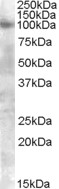
Images (1) Click the Picture to Zoom In
-
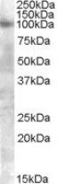
ARG64673 anti-FGFR2 antibody WB image
Western Blot: A549 lysate (35 µg protein in RIPA buffer) stained with ARG64673 anti-FGFR2 antibody at 0.3 µg/ml dilution.