ARG63738
anti-FGFR1 antibody
anti-FGFR1 antibody for IHC-Formalin-fixed paraffin-embedded sections,Western blot and Human
Cancer antibody; Cell Biology and Cellular Response antibody; Gene Regulation antibody; Neuroscience antibody; Signaling Transduction antibody
Overview
| Product Description | Goat Polyclonal antibody recognizes FGFR1 |
|---|---|
| Tested Reactivity | Hu |
| Predict Reactivity | Cow, Dog |
| Tested Application | IHC-P, WB |
| Specificity | This antibody is expected to recognise five isoforms (as represented by NP_075598.2; NP_056934.2; NP_075593.1; NP_075594.1; NP_075599.1). |
| Host | Goat |
| Clonality | Polyclonal |
| Isotype | IgG |
| Target Name | FGFR1 |
| Antigen Species | Human |
| Immunogen | CLPRHPAQLANGGLKR |
| Conjugation | Un-conjugated |
| Alternate Names | HH2; bFGF-R-1; Fibroblast growth factor receptor 1; Proto-oncogene c-Fgr; FLT2; FLG; CD antigen CD331; CEK; N-SAM; Fms-like tyrosine kinase 2; HBGFR; Basic fibroblast growth factor receptor 1; N-sam; CD331; FLT-2; FGFR-1; FGFBR; BFGFR; HRTFDS; KAL2; OGD; EC 2.7.10.1 |
Application Instructions
| Application Suggestion |
|
||||||
|---|---|---|---|---|---|---|---|
| Application Note | WB: Recommend incubate at RT for 1h. IHC-P: Antigen Retrieval: Steam tissue section in Citrate buffer (pH 6.0). * The dilutions indicate recommended starting dilutions and the optimal dilutions or concentrations should be determined by the scientist. |
Properties
| Form | Liquid |
|---|---|
| Purification | Purified from goat serum by antigen affinity chromatography. |
| Buffer | Tris saline (pH 7.3), 0.02% Sodium azide and 0.5% BSA. |
| Preservative | 0.02% Sodium azide |
| Stabilizer | 0.5% BSA |
| Concentration | 0.5 mg/ml |
| Storage Instruction | For continuous use, store undiluted antibody at 2-8°C for up to a week. For long-term storage, aliquot and store at -20°C or below. Storage in frost free freezers is not recommended. Avoid repeated freeze/thaw cycles. Suggest spin the vial prior to opening. The antibody solution should be gently mixed before use. |
| Note | For laboratory research only, not for drug, diagnostic or other use. |
Bioinformation
| Database Links |
Swiss-port # P11362 Human Fibroblast growth factor receptor 1 |
|---|---|
| Background | The protein encoded by this gene is a member of the fibroblast growth factor receptor (FGFR) family, where amino acid sequence is highly conserved between members and throughout evolution. FGFR family members differ from one another in their ligand affinities and tissue distribution. A full-length representative protein consists of an extracellular region, composed of three immunoglobulin-like domains, a single hydrophobic membrane-spanning segment and a cytoplasmic tyrosine kinase domain. The extracellular portion of the protein interacts with fibroblast growth factors, setting in motion a cascade of downstream signals, ultimately influencing mitogenesis and differentiation. This particular family member binds both acidic and basic fibroblast growth factors and is involved in limb induction. Mutations in this gene have been associated with Pfeiffer syndrome, Jackson-Weiss syndrome, Antley-Bixler syndrome, osteoglophonic dysplasia, and autosomal dominant Kallmann syndrome 2. Chromosomal aberrations involving this gene are associated with stem cell myeloproliferative disorder and stem cell leukemia lymphoma syndrome. Alternatively spliced variants which encode different protein isoforms have been described; however, not all variants have been fully characterized. [provided by RefSeq, Jul 2008] |
| Research Area | Cancer antibody; Cell Biology and Cellular Response antibody; Gene Regulation antibody; Neuroscience antibody; Signaling Transduction antibody |
| Calculated MW | 92 kDa |
| PTM | Autophosphorylated. Binding of FGF family members together with heparan sulfate proteoglycan or heparin promotes receptor dimerization and autophosphorylation on tyrosine residues. Autophosphorylation occurs in trans between the two FGFR molecules present in the dimer and proceeds in a highly ordered manner. Initial autophosphorylation at Tyr-653 increases the kinase activity by a factor of 50 to 100. After this, Tyr-583 becomes phosphorylated, followed by phosphorylation of Tyr-463, Tyr-766, Tyr-583 and Tyr-585. In a third stage, Tyr-654 is autophosphorylated, resulting in a further tenfold increase of kinase activity. Phosphotyrosine residues provide docking sites for interacting proteins and so are crucial for FGFR1 function and its regulation. Ubiquitinated. FGFR1 is rapidly ubiquitinated by NEDD4 after autophosphorylation, leading to internalization and lysosomal degradation. CBL is recruited to activated FGFR1 via FRS2 and GRB2, and mediates ubiquitination and subsequent degradation of FGFR1. N-glycosylated in the endoplasmic reticulum. The N-glycan chains undergo further maturation to an Endo H-resistant form in the Golgi apparatus. |
Images (3) Click the Picture to Zoom In
-

ARG63738 anti-FGFR1 antibody WB image
Western Blot: human breast lysate (35 µg protein in RIPA buffer) stained with ARG63738 anti-FGFR1 antibody at 0.3 µg/ml dilution.
-
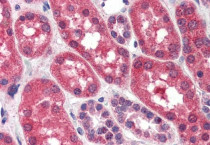
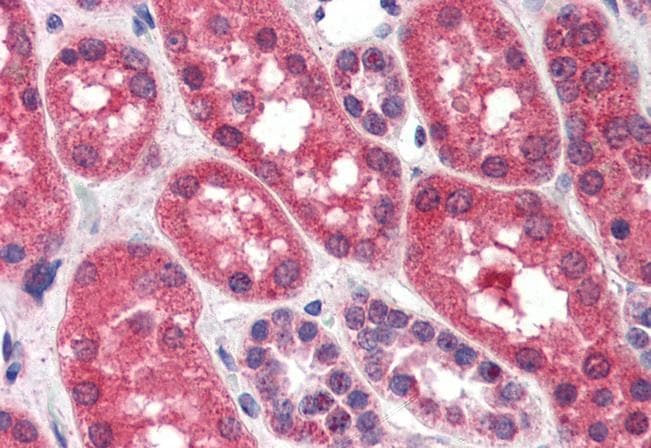
ARG63738 anti-FGFR1 antibody IHC-P image
Immunohistochemistry: Paraffin-embedded Human kidney tissue. Antigen Retrieval: Steam tissue section in Citrate buffer (pH 6.0). The tissue section was stained with ARG63738 anti-FGFR1 antibody at 4 µg/ml dilution followed by AP-staining.
-
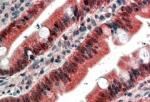
ARG63738 anti-FGFR1 antibody IHC-P image
Immunohistochemistry: paraffin embedded Human Small Intestine. (Steamed antigen retrieval with citrate buffer pH 6) stained with ARG63738 anti-FGFR1 antibody at 4 µg/ml dilution followed by AP-staining.