ARG66701
anti-FAP / Fibroblast activation protein antibody
anti-FAP / Fibroblast activation protein antibody for ICC/IF,IHC-Formalin-fixed paraffin-embedded sections,Western blot and Human,Mouse,Rat
Overview
| Product Description | Rabbit Polyclonal antibody recognizes FAP / Fibroblast activation protein |
|---|---|
| Tested Reactivity | Hu, Ms, Rat |
| Tested Application | ICC/IF, IHC-P, WB |
| Host | Rabbit |
| Clonality | Polyclonal |
| Isotype | IgG |
| Target Name | FAP / Fibroblast activation protein |
| Antigen Species | Human |
| Immunogen | KLH-conjugated synthetic peptide within the center region of Human FAP / Fibroblast activation protein. |
| Conjugation | Un-conjugated |
| Alternate Names | Fibroblast activation protein alpha; SIMP; Seprase; Integral membrane serine protease; Surface-expressed protease; EC 3.4.21.26; Dipeptidyl peptidase FAP; 170 kDa melanoma membrane-bound gelatinase; EC 3.4.14.5; APCE; Serine integral membrane protease; DPPIV; EC 3.4.21.-; FAPalpha; Gelatine degradation protease FAP; Post-proline cleaving enzyme; Prolyl endopeptidase FAP; FAPA |
Application Instructions
| Application Suggestion |
|
||||||||
|---|---|---|---|---|---|---|---|---|---|
| Application Note | IHC-P: Antigen Retrieval: Heat mediation was performed in Sodium citrate buffer (pH 6.0). * The dilutions indicate recommended starting dilutions and the optimal dilutions or concentrations should be determined by the scientist. |
||||||||
| Observed Size | ~ 88 kDa |
Properties
| Form | Liquid |
|---|---|
| Purification | Affinity purification with immunogen. |
| Buffer | 0.42% Potassium phosphate (pH 7.3), 0.87% NaCl, 0.01% Sodium azide and 30% Glycerol. |
| Preservative | 0.01% Sodium azide |
| Stabilizer | 30% Glycerol |
| Storage Instruction | For continuous use, store undiluted antibody at 2-8°C for up to a week. For long-term storage, aliquot and store at -20°C. Storage in frost free freezers is not recommended. Avoid repeated freeze/thaw cycles. Suggest spin the vial prior to opening. The antibody solution should be gently mixed before use. |
| Note | For laboratory research only, not for drug, diagnostic or other use. |
Bioinformation
| Database Links | |
|---|---|
| Gene Symbol | FAP |
| Gene Full Name | fibroblast activation protein, alpha |
| Background | The protein encoded by this gene is a homodimeric integral membrane gelatinase belonging to the serine protease family. It is selectively expressed in reactive stromal fibroblasts of epithelial cancers, granulation tissue of healing wounds, and malignant cells of bone and soft tissue sarcomas. This protein is thought to be involved in the control of fibroblast growth or epithelial-mesenchymal interactions during development, tissue repair, and epithelial carcinogenesis. Alternatively spliced transcript variants encoding different isoforms have been found for this gene. [provided by RefSeq, Apr 2014] |
| Function | Cell surface glycoprotein serine protease that participates in extracellular matrix degradation and involved in many cellular processes including tissue remodeling, fibrosis, wound healing, inflammation and tumor growth. Both plasma membrane and soluble forms exhibit post-proline cleaving endopeptidase activity, with a marked preference for Ala/Ser-Gly-Pro-Ser/Asn/Ala consensus sequences, on substrate such as alpha-2-antiplasmin SERPINF2 and SPRY2. Degrade also gelatin, heat-denatured type I collagen, but not native collagen type I and IV, vibronectin, tenascin, laminin, fibronectin, fibrin or casein. Have also dipeptidyl peptidase activity, exhibiting the ability to hydrolyze the prolyl bond two residues from the N-terminus of synthetic dipeptide substrates provided that the penultimate residue is proline, with a preference for Ala-Pro, Ile-Pro, Gly-Pro, Arg-Pro and Pro-Pro. Natural neuropeptide hormones for dipeptidyl peptidase are the neuropeptide Y (NPY), peptide YY (PYY), substance P (TAC1) and brain natriuretic peptide 32 (NPPB). The plasma membrane form, in association with either DPP4, PLAUR or integrins, is involved in the pericellular proteolysis of the extracellular matrix (ECM), and hence promotes cell adhesion, migration and invasion through the ECM. Plays a role in tissue remodeling during development and wound healing. Participates in the cell invasiveness towards the ECM in malignant melanoma cancers. Enhances tumor growth progression by increasing angiogenesis, collagen fiber degradation and apoptosis and by reducing antitumor response of the immune system. Promotes glioma cell invasion through the brain parenchyma by degrading the proteoglycan brevican. Acts as a tumor suppressor in melanocytic cells through regulation of cell proliferation and survival in a serine protease activity-independent manner. [UniProt] |
| Cellular Localization | Plasma membrane. Note: Localized on cell surface with lamellipodia and invadopodia membranes and on shed vesicles. Secreted. Note: Found in blood plasma and serum. [UniProt] |
| Highlight | Related Antibody Duos and Panels: ARG30347 CAF Marker Antibody Panel Related products: Fibroblast activation protein antibodies; Fibroblast activation protein ELISA Kits; Fibroblast activation protein Duos / Panels; Anti-Rabbit IgG secondary antibodies; Related news: New antibody panels for Myofibroblasts and CAFs |
| Calculated MW | 88 kDa |
| PTM | N-glycosylated. The N-terminus may be blocked. [UniProt] |
Images (3) Click the Picture to Zoom In
-
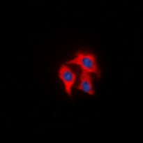
ARG66701 anti-FAP / Fibroblast activation protein antibody ICC/IF image
Immunofluorescence: Formalin-fixed HeLa cells were permeabilized with 0.1% Triton X-100 in TBS for 5-10 minutes and blocked with 3% BSA-PBS for 30 minutes at room temperature. Cells were stained with ARG66701 anti-FAP / Fibroblast activation protein antibody (red) in 3% BSA-PBS and incubated overnight at 4°C. DAPI (blue) for nuclear staining.
-
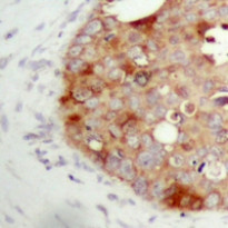
ARG66701 anti-FAP / Fibroblast activation protein antibody IHC-P image
Immunohistochemistry: Formalin-fixed and paraffin-embedded Human prostate cancer tissue. Antigen Retrieval: Heat mediation was performed in Sodium citrate buffer (pH 6.0). The tissue section was stained with ARG66701 anti-FAP / Fibroblast activation protein antibody at room temperature. The section was counterstained with haematoxylin and mounted with DPX.
-
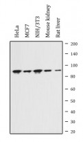
ARG66701 anti-FAP / Fibroblast activation protein antibody WB image
Western blot: HeLa, MCF7, NIH/3T3, Mouse kidney and Rat liver lysates stained with ARG66701 anti-FAP / Fibroblast activation protein antibody.


Specific References
Exploring the Role of Fibroblasts in Promoting Neuroblastoma Cell Migration and Invasion
ICC/IF / Human


