ARG58618
anti-Ephrin A1 antibody
anti-Ephrin A1 antibody for Western blot and Mouse,Rat
Overview
| Product Description | Goat Polyclonal antibody recognizes Ephrin A1 |
|---|---|
| Tested Reactivity | Ms, Rat |
| Predict Reactivity | Hu, Dog, Pig |
| Tested Application | WB |
| Host | Goat |
| Clonality | Polyclonal |
| Isotype | IgG |
| Target Name | Ephrin A1 |
| Antigen Species | Human |
| Immunogen | Synthetic peptide from the internal region of Human Ephrin A1 (NP_004419.2). (C-KVTVSGKITHSP) |
| Conjugation | Un-conjugated |
| Alternate Names | ECKLG; B61; EPLG1; TNFAIP4; Immediate early response protein B61; Tumor necrosis factor alpha-induced protein 4; EFL1; LERK-1; EPH-related receptor tyrosine kinase ligand 1; LERK1; TNF alpha-induced protein 4; Ephrin-A1 |
Application Instructions
| Application Suggestion |
|
||||
|---|---|---|---|---|---|
| Application Note | WB: Recommend incubate at RT for 1h. * The dilutions indicate recommended starting dilutions and the optimal dilutions or concentrations should be determined by the scientist. |
||||
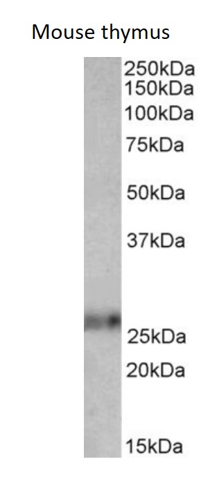
| Observed Size | ~ 26 kDa |
Properties
| Form | Liquid |
|---|---|
| Purification | Affinity purified |
| Buffer | Tris saline (pH 7.3), 0.02% Sodium azide and 0.5% BSA. |
| Preservative | 0.02% Sodium azide |
| Stabilizer | 0.5% BSA |
| Concentration | 0.5 mg/ml |
| Storage Instruction | For continuous use, store undiluted antibody at 2-8°C for up to a week. For long-term storage, aliquot and store at -20°C or below. Storage in frost free freezers is not recommended. Avoid repeated freeze/thaw cycles. Suggest spin the vial prior to opening. The antibody solution should be gently mixed before use. |
| Note | For laboratory research only, not for drug, diagnostic or other use. |
Bioinformation
| Database Links | |
|---|---|
| Gene Symbol | EFNA1 |
| Gene Full Name | ephrin-A1 |
| Background | This gene encodes a member of the ephrin (EPH) family. The ephrins and EPH-related receptors comprise the largest subfamily of receptor protein-tyrosine kinases and have been implicated in mediating developmental events, especially in the nervous system and in erythropoiesis. Based on their structures and sequence relationships, ephrins are divided into the ephrin-A (EFNA) class, which are anchored to the membrane by a glycosylphosphatidylinositol linkage, and the ephrin-B (EFNB) class, which are transmembrane proteins. This gene encodes an EFNA class ephrin which binds to the EPHA2, EPHA4, EPHA5, EPHA6, and EPHA7 receptors. Two transcript variants that encode different isoforms were identified through sequence analysis. [provided by RefSeq, Jul 2008] |
| Function | Cell surface GPI-bound ligand for Eph receptors, a family of receptor tyrosine kinases which are crucial for migration, repulsion and adhesion during neuronal, vascular and epithelial development. Binds promiscuously Eph receptors residing on adjacent cells, leading to contact-dependent bidirectional signaling into neighboring cells. Plays an important role in angiogenesis and tumor neovascularization. The recruitment of VAV2, VAV3 and PI3-kinase p85 subunit by phosphorylated EPHA2 is critical for EFNA1-induced RAC1 GTPase activation and vascular endothelial cell migration and assembly. Exerts anti-oncogenic effects in tumor cells through activation and down-regulation of EPHA2. Activates EPHA2 by inducing tyrosine phosphorylation which leads to its internalization and degradation. Acts as a negative regulator in the tumorigenesis of gliomas by down-regulating EPHA2 and FAK. Can evoke collapse of embryonic neuronal growth cone and regulates dendritic spine morphogenesis. [UniProt] |
| Calculated MW | 24 kDa |
| PTM | Undergoes proteolysis by a metalloprotease to give rise to a soluble monomeric form. N-Glycosylation is required for binding to EPHA2 receptor and inducing its internalization. [UniProt] |
Images (1) Click the Picture to Zoom In
-
ARG58618 anti-Ephrin A1 antibody WB image
Western blot: 35 µg of Mouse thymus lysate (in RIPA buffer) stained with ARG58618 anti-Ephrin A1 antibody at 0.01 µg/ml dilution. Primary incubation was 1 hour. Detected by chemiluminescence.