ARG42066
anti-EphA8 antibody
anti-EphA8 antibody for Western blot and Human,Mouse,Rat
Overview
| Product Description | Rabbit Polyclonal antibody recognizes EphA8 |
|---|---|
| Tested Reactivity | Hu, Ms, Rat |
| Tested Application | WB |
| Host | Rabbit |
| Clonality | Polyclonal |
| Isotype | IgG |
| Target Name | EphA8 |
| Antigen Species | Human |
| Immunogen | Synthetic peptide of Human EphA8. |
| Conjugation | Un-conjugated |
| Alternate Names | EPH- and ELK-related kinase; EK3; hEK3; EPH-like kinase 3; HEK3; Tyrosine-protein kinase receptor EEK; EEK; Ephrin type-A receptor 8; EC 2.7.10.1 |
Application Instructions
| Application Suggestion |
|
||||
|---|---|---|---|---|---|
| Application Note | * The dilutions indicate recommended starting dilutions and the optimal dilutions or concentrations should be determined by the scientist. | ||||
| Observed Size | ~ 111 kDa - 120 kDa |
Properties
| Form | Liquid |
|---|---|
| Purification | Affinity purification with immunogen. |
| Purity | > 95% (by SDS-PAGE). |
| Buffer | PBS (pH 7.2), 0.02% Sodium azide and 50% Glycerol. |
| Preservative | 0.02% Sodium azide |
| Stabilizer | 50% Glycerol |
| Concentration | 1 mg/ml |
| Storage Instruction | For continuous use, store undiluted antibody at 2-8°C for up to a week. For long-term storage, aliquot and store at -20°C. Storage in frost free freezers is not recommended. Avoid repeated freeze/thaw cycles. Suggest spin the vial prior to opening. The antibody solution should be gently mixed before use. |
| Note | For laboratory research only, not for drug, diagnostic or other use. |
Bioinformation
| Database Links | |
|---|---|
| Gene Symbol | EPHA8 |
| Gene Full Name | EPH receptor A8 |
| Background | This gene encodes a member of the ephrin receptor subfamily of the protein-tyrosine kinase family. EPH and EPH-related receptors have been implicated in mediating developmental events, particularly in the nervous system. Receptors in the EPH subfamily typically have a single kinase domain and an extracellular region containing a Cys-rich domain and 2 fibronectin type III repeats. The ephrin receptors are divided into 2 groups based on the similarity of their extracellular domain sequences and their affinities for binding ephrin-A and ephrin-B ligands. The protein encoded by this gene functions as a receptor for ephrin A2, A3 and A5 and plays a role in short-range contact-mediated axonal guidance during development of the mammalian nervous system. [provided by RefSeq, Jul 2008] |
| Function | Receptor tyrosine kinase which binds promiscuously GPI-anchored ephrin-A family ligands residing on adjacent cells, leading to contact-dependent bidirectional signaling into neighboring cells. The signaling pathway downstream of the receptor is referred to as forward signaling while the signaling pathway downstream of the ephrin ligand is referred to as reverse signaling. The GPI-anchored ephrin-A EFNA2, EFNA3, and EFNA5 are able to activate EPHA8 through phosphorylation. With EFNA5 may regulate integrin-mediated cell adhesion and migration on fibronectin substrate but also neurite outgrowth. During development of the nervous system plays also a role in axon guidance. Downstream effectors of the EPHA8 signaling pathway include FYN which promotes cell adhesion upon activation by EPHA8 and the MAP kinases in the stimulation of neurite outgrowth (By similarity). [UniProt] |
| Cellular Localization | Cell membrane; Single-pass type I membrane protein. Cell projection. Early endosome membrane. Note=Undergoes clathrin-mediated endocytosis upon EFNA5-binding and is targeted to early endosomes. [UniProt] |
| Calculated MW | 111 kDa |
| PTM | Phosphorylated. Phosphorylation is stimulated upon binding of its ligands including EFNA2, EFNA3 and EFNA5. Autophosphorylation on Tyr-616 is critical for association with FYN. Autophosphorylation on Tyr-839 modulates tyrosine kinase activity (By similarity). Ubiquitinated. Ubiquitination by CBL regulates the receptor stability and activity through proteasomal degradation. ANKS1A prevents ubiquitination and degradation (By similarity). [UniProt] |
Images (2) Click the Picture to Zoom In
-

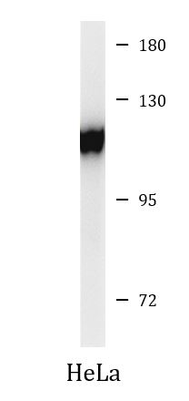
ARG42066 anti-EphA8 antibody WB image
Western blot: 40 µg of HeLa whole cell lysate stained with ARG42066 anti-EphA8 antibody at 1:500 dilution.
-
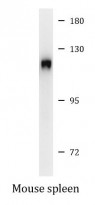
ARG42066 anti-EphA8 antibody WB image
Western blot: 40 µg of Mouse spleen lysate stained with ARG42066 anti-EphA8 antibody at 1:500 dilution.