ARG55758
anti-EphA4 antibody
anti-EphA4 antibody for IHC-Formalin-fixed paraffin-embedded sections,Western blot and Human,Mouse
Overview
| Product Description | Rabbit Polyclonal antibody recognizes EphA4 |
|---|---|
| Tested Reactivity | Hu, Ms |
| Tested Application | IHC-P, WB |
| Host | Rabbit |
| Clonality | Polyclonal |
| Isotype | IgG |
| Target Name | EphA4 |
| Antigen Species | Human |
| Immunogen | KLH-conjugated synthetic peptide corresponding to aa. 875-904 (C-terminus) of Human EphA4. |
| Conjugation | Un-conjugated |
| Alternate Names | EC 2.7.10.1; Tyrosine-protein kinase receptor SEK; HEK8; SEK; hEK8; Tyrosine-protein kinase TYRO1; Ephrin type-A receptor 4; EK8; EPH-like kinase 8; TYRO1 |
Application Instructions
| Application Suggestion |
|
||||||
|---|---|---|---|---|---|---|---|
| Application Note | * The dilutions indicate recommended starting dilutions and the optimal dilutions or concentrations should be determined by the scientist. | ||||||
| Positive Control | NIH/3T3 |
Properties
| Form | Liquid |
|---|---|
| Purification | Purification with Protein G. |
| Buffer | PBS and 0.09% (W/V) Sodium azide. |
| Preservative | 0.09% (W/V) Sodium azide. |
| Storage Instruction | For continuous use, store undiluted antibody at 2-8°C for up to a week. For long-term storage, aliquot and store at -20°C or below. Storage in frost free freezers is not recommended. Avoid repeated freeze/thaw cycles. Suggest spin the vial prior to opening. The antibody solution should be gently mixed before use. |
| Note | For laboratory research only, not for drug, diagnostic or other use. |
Bioinformation
| Database Links | |
|---|---|
| Gene Symbol | EPHA4 |
| Gene Full Name | EPH receptor A4 |
| Background | This gene belongs to the ephrin receptor subfamily of the protein-tyrosine kinase family. EPH and EPH-related receptors have been implicated in mediating developmental events, particularly in the nervous system. Receptors in the EPH subfamily typically have a single kinase domain and an extracellular region containing a Cys-rich domain and 2 fibronectin type III repeats. The ephrin receptors are divided into 2 groups based on the similarity of their extracellular domain sequences and their affinities for binding ephrin-A and ephrin-B ligands. Multiple transcript variants encoding different isoforms have been found for this gene. [provided by RefSeq, Jan 2015] |
| Function | Receptor tyrosine kinase which binds membrane-bound ephrin family ligands residing on adjacent cells, leading to contact-dependent bidirectional signaling into neighboring cells. The signaling pathway downstream of the receptor is referred to as forward signaling while the signaling pathway downstream of the ephrin ligand is referred to as reverse signaling. Highly promiscuous, it has the unique property among Eph receptors to bind and to be physiologically activated by both GPI-anchored ephrin-A and transmembrane ephrin-B ligands including EFNA1 and EFNB3. Upon activation by ephrin ligands, modulates cell morphology and integrin-dependent cell adhesion through regulation of the Rac, Rap and Rho GTPases activity. Plays an important role in the development of the nervous system controlling different steps of axonal guidance including the establishment of the corticospinal projections. May also control the segregation of motor and sensory axons during neuromuscular circuit development. In addition to its role in axonal guidance plays a role in synaptic plasticity. Activated by EFNA1 phosphorylates CDK5 at 'Tyr-15' which in turn phosphorylates NGEF regulating RHOA and dendritic spine morphogenesis. In the nervous system, plays also a role in repair after injury preventing axonal regeneration and in angiogenesis playing a role in central nervous system vascular formation. Additionally, its promiscuity makes it available to participate in a variety of cell-cell signaling regulating for instance the development of the thymic epithelium. [UniProt] |
| Cellular Localization | Cell membrane; Single-pass type I membrane protein. Cell projection, axon. Cell projection, dendrite. Cell junction, synapse, postsynaptic cell membrane, postsynaptic density. Early endosome Note=Clustered upon activation and targeted to early endosome |
| Calculated MW | 110 kDa |
Images (2) Click the Picture to Zoom In
-

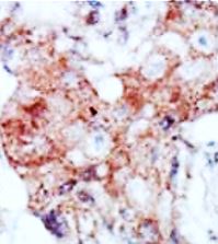
ARG55758 anti-EphA4 antibody IHC-P image
Immunohistochemistry: Formalin-fixed and paraffin-embedded Human breast carcinoma tissue stained with ARG55758 anti-EphA4 antibody.
-

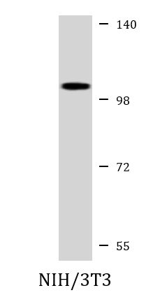
ARG55758 anti-EphA4 antibody WB image
Western blot: 35 µg of NIH/3T3 cell lysate stained with ARG55758 anti-EphA4 antibody at 1:1000 dilution.