ARG43755
anti-EpCAM antibody
anti-EpCAM antibody for Flow cytometry,IHC-Formalin-fixed paraffin-embedded sections and Human
Controls and Markers antibody; Epithelial Marker antibody; Circulating Tumor Cells BioMarker antibody
Overview
| Product Description | Rabbit Polyclonal antibody recognizes EpCAM |
|---|---|
| Tested Reactivity | Hu |
| Tested Application | FACS, IHC-P |
| Host | Rabbit |
| Clonality | Polyclonal |
| Isotype | IgG |
| Target Name | EpCAM |
| Antigen Species | Human |
| Immunogen | Synthetic peptide corresponding to a.a. E147-N189 in extracellular domain of Human EpCAM. |
| Conjugation | Un-conjugated |
| Alternate Names | MIC18; EGP; Tumor-associated calcium signal transducer 1; Epithelial glycoprotein 314; KSA; Ep-CAM; Epithelial cell surface antigen; Adenocarcinoma-associated antigen; HNPCC8; Cell surface glycoprotein Trop-1; EGP40; TACSTD1; KS1/4; hEGP314; Major gastrointestinal tumor-associated protein GA733-2; M4S1; MK-1; Epithelial glycoprotein; KS 1/4 antigen; ESA; DIAR5; EGP314; Epithelial cell adhesion molecule; EGP-2; TROP1; CD antigen CD326 |
Application Instructions
| Application Suggestion |
|
||||||
|---|---|---|---|---|---|---|---|
| Application Note | * The dilutions indicate recommended starting dilutions and the optimal dilutions or concentrations should be determined by the scientist. | ||||||
| Observed Size | 40 kDa |
Properties
| Form | Liquid |
|---|---|
| Purification | Affinity purification with immunogen. |
| Buffer | 0.9% NaCl, 0.2% Na2HPO4, 0.05% Sodium azide and 5% BSA. |
| Preservative | 0.05% Sodium azide |
| Stabilizer | 5% BSA |
| Concentration | 0.5 mg/ml |
| Storage Instruction | For continuous use, store undiluted antibody at 2-8°C for up to a week. For long-term storage, aliquot and store at -20°C. Storage in frost free freezers is not recommended. Avoid repeated freeze/thaw cycles. Suggest spin the vial prior to opening. The antibody solution should be gently mixed before use. |
| Note | For laboratory research only, not for drug, diagnostic or other use. |
Bioinformation
| Database Links | |
|---|---|
| Gene Symbol | EPCAM |
| Gene Full Name | epithelial cell adhesion molecule |
| Background | EpCAM is a carcinoma-associated antigen and is a member of a family that includes at least two type I membrane proteins. This antigen is expressed on most normal epithelial cells and gastrointestinal carcinomas and functions as a homotypic calcium-independent cell adhesion molecule. The antigen is being used as a target for immunotherapy treatment of human carcinomas. Mutations in this gene result in congenital tufting enteropathy. [provided by RefSeq, Dec 2008] |
| Function | EpCAM may act as a physical homophilic interaction molecule between intestinal epithelial cells (IECs) and intraepithelial lymphocytes (IELs) at the mucosal epithelium for providing immunological barrier as a first line of defense against mucosal infection. Plays a role in embryonic stem cells proliferation and differentiation. Up-regulates the expression of FABP5, MYC and cyclins A and E. [UniProt] |
| Cellular Localization | Lateral cell membrane; Single-pass type I membrane protein. Cell junction, tight junction. Note=Colocalizes with CLDN7 at the lateral cell membrane and tight junction. [UniProt] |
| Research Area | Controls and Markers antibody; Epithelial Marker antibody; Circulating Tumor Cells BioMarker antibody |
| Calculated MW | 35 kDa |
| PTM | Hyperglycosylated in carcinoma tissue as compared with autologous normal epithelia. Glycosylation at Asn-198 is crucial for protein stability. [UniProt] |
Images (5) Click the Picture to Zoom In
-
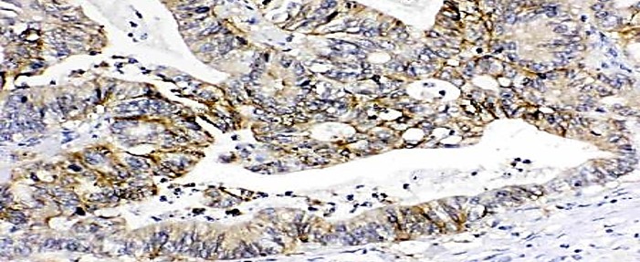
ARG43755 anti-EpCAM antibody IHC-P image
Immunohistochemistry: Paraffin-embedded Human intestinal cancer tissue. Antigen Retrieval: Heat mediation was performed in EDTA buffer (pH 8.0). The tissue section was blocked with 10% goat serum. The tissue section was then stained with ARG43755 anti-EpCAM antibody at 1 µg/ml dilution, overnight at 4°C.
-
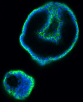
ARG43755 anti-EpCAM antibody ICC/IF image
Immunofluorescence: Human colon organoid cells were stained with ARG43755 anti-EpCAM antibody (green) at 1:200 and 4°C. DAPI (blue) was used as the nuclear counter stain.Antigen Retrieval: Heat mediated was performed using citrate buffer pH 6.0.
-
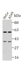
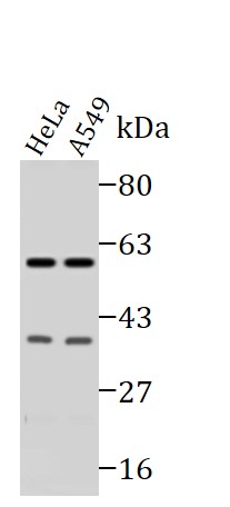
ARG43755 anti-EpCAM antibody WB image
Western blot: 10 µg of HeLa and A549 cell lysates stained with ARG43755 anti-EpCAM antibody at 1:1000 dilution
-
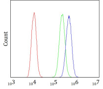
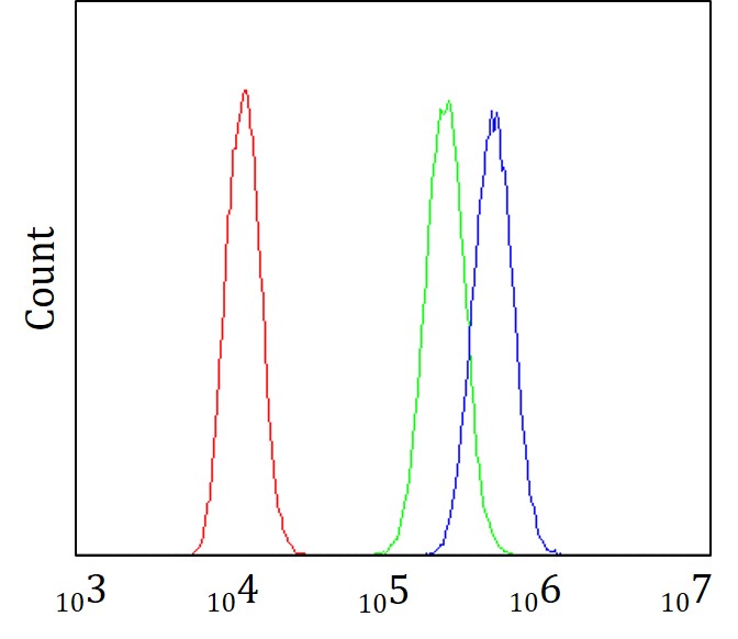
ARG43755 anti-EpCAM antibody FACS image
Flow Cytometry: A431 cells were blocked with 10% normal goat serum and stained with ARG43755 anti-EpCAM antibody (blue) at 1 µg/10^6 cells for 30 min at 20°C, followed by incubation with DyLight®488 labelled secondary antibody. Isotype control antibody (green) was rabbit IgG (1 µg/10^6 cells) used under the same conditions. Unlabelled sample (red) was also used as a control.
-
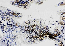
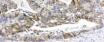
ARG43755 anti-EpCAM antibody IHC-P image
Immunohistochemistry: Paraffin-embedded Human prostatic cancer tissue. Antigen Retrieval: Heat mediation was performed in EDTA buffer (pH 8.0). The tissue section was blocked with 10% goat serum. The tissue section was then stained with ARG43755 anti-EpCAM antibody at 1 µg/ml dilution, overnight at 4°C.