ARG55159
anti-Enolase 1 antibody
anti-Enolase 1 antibody for Flow cytometry,ICC/IF,IHC-Formalin-fixed paraffin-embedded sections,Western blot and Human,Mouse,Rat
Cancer antibody; Gene Regulation antibody; Metabolism antibody; Signaling Transduction antibody

Overview
| Product Description | Rabbit Polyclonal antibody recognizes Enolase 1 |
|---|---|
| Tested Reactivity | Hu, Ms, Rat |
| Predict Reactivity | Bov, Rat, Chk, Mk, Xenopus |
| Tested Application | FACS, ICC/IF, IHC-P, WB |
| Host | Rabbit |
| Clonality | Polyclonal |
| Isotype | IgG |
| Target Name | Enolase 1 |
| Antigen Species | Human |
| Immunogen | KLH-conjugated synthetic peptide corresponding to aa. 178-205 (Center) of Human Enolase 1. |
| Conjugation | Un-conjugated |
| Alternate Names | MPB1; Plasminogen-binding protein; Alpha-enolase; MBP-1; NNE; PPH; Enolase 1; ENO1L1; Phosphopyruvate hydratase; 2-phospho-D-glycerate hydro-lyase; C-myc promoter-binding protein; Non-neural enolase; MPB-1; EC 4.2.1.11 |
Application Instructions
| Application Suggestion |
|
||||||||||
|---|---|---|---|---|---|---|---|---|---|---|---|
| Application Note | * The dilutions indicate recommended starting dilutions and the optimal dilutions or concentrations should be determined by the scientist. |
Properties
| Form | Liquid |
|---|---|
| Purification | Saturated Ammonium Sulfate (SAS) precipitation followed by dialysis against PBS. |
| Buffer | PBS and 0.09% (W/V) Sodium azide |
| Preservative | 0.09% (W/V) Sodium azide |
| Storage Instruction | For continuous use, store undiluted antibody at 2-8°C for up to a week. For long-term storage, aliquot and store at -20°C or below. Storage in frost free freezers is not recommended. Avoid repeated freeze/thaw cycles. Suggest spin the vial prior to opening. The antibody solution should be gently mixed before use. |
| Note | For laboratory research only, not for drug, diagnostic or other use. |
Bioinformation
| Database Links | |
|---|---|
| Gene Symbol | ENO1 |
| Gene Full Name | enolase 1, (alpha) |
| Background | This gene encodes alpha-enolase, one of three enolase isoenzymes found in mammals. Each isoenzyme is a homodimer composed of 2 alpha, 2 gamma, or 2 beta subunits, and functions as a glycolytic enzyme. Alpha-enolase in addition, functions as a structural lens protein (tau-crystallin) in the monomeric form. Alternative splicing of this gene results in a shorter isoform that has been shown to bind to the c-myc promoter and function as a tumor suppressor. Several pseudogenes have been identified, including one on the long arm of chromosome 1. Alpha-enolase has also been identified as an autoantigen in Hashimoto encephalopathy. [provided by RefSeq, Jan 2011] |
| Function | Multifunctional enzyme that, as well as its role in glycolysis, plays a part in various processes such as growth control, hypoxia tolerance and allergic responses. May also function in the intravascular and pericellular fibrinolytic system due to its ability to serve as a receptor and activator of plasminogen on the cell surface of several cell-types such as leukocytes and neurons. Stimulates immunoglobulin production. MBP1 binds to the myc promoter and acts as a transcriptional repressor. May be a tumor suppressor. [UniProt] |
| Cellular Localization | Cytoplasm. Cell membrane. Cytoplasm, myofibril, sarcomere, M line. Note=Can translocate to the plasma membrane in either the homodimeric (alpha/alpha) or heterodimeric (alpha/gamma) form. ENO1 is localized to the M line |
| Research Area | Cancer antibody; Gene Regulation antibody; Metabolism antibody; Signaling Transduction antibody |
| Calculated MW | 47 kDa |
| PTM | ISGylated. |
Images (4) Click the Picture to Zoom In
-
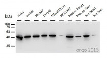
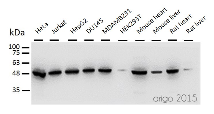
ARG55159 anti-Enolase 1 antibody WB image
Western blot: 30 µg of HeLa, Jurkat , HepG2, DU145, MDAMB231, HEK293T, Mouse heart, Mouse liver, Rat heart and Rat liver lysates stained with ARG55159 anti-Enolase 1 antibody at 1:500 dilution.
-
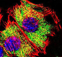
ARG55159 anti-Enolase 1 antibody ICC/IF image
Immunofluorescence: C2C12 cells were fixed with 4% PFA (20 min), permeabilized with Triton X-100 (0.1%, 10 min), then stained with ARG55159 anti-Enolase 1 antibody (green) at 1:25 dilution, 1 hour at 37°C. Cytoplasmic actin was counterstained with Alexa Fluor® 555 (red) conjugated Phalloidin (7 units/ml, 1 hour at 37°C). Nuclei were counterstained with DAPI (blue) (10 µg/ml, 10 min).
-

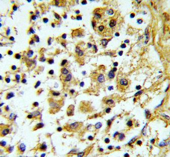
ARG55159 anti-Enolase 1 antibody IHC-P image
Immunohistochemistry: Formalin-fixed and paraffin-embedded Human lymph stained with ARG55159 anti-Enolase 1 antibody.
-
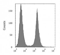
ARG55159 anti-Enolase 1 antibody FACS image
Flow Cytometry: HeLa cells stained with ARG55159 anti-Enolase 1 antibody (right histogram) or without primary antibody control (left histogram), followed by incubation with FITC labelled secondary antibody.


Customer's Feedback
Excellent

Review for anti-Enolase 1 antibody
Application:WB
Sample:MCF-7
Sample Loading Amount:30 µg
Primary Antibody Dilution Factor:1:500
Primary Antibody Incubation Time:overnight
Primary Antibody Incubation Temperature:4 ºC


