ARG58617
anti-ERV3 antibody
anti-ERV3 antibody for IHC-Formalin-fixed paraffin-embedded sections,Western blot and Human
Overview
| Product Description | Goat Polyclonal antibody recognizes ERV3 |
|---|---|
| Tested Reactivity | Hu |
| Tested Application | IHC-P, WB |
| Host | Goat |
| Clonality | Polyclonal |
| Isotype | IgG |
| Target Name | ERV3 |
| Antigen Species | Human |
| Immunogen | Synthetic peptide from the internal region of Human ERV3 (NP_001007254.2). (C-PQDNFTLTASS) |
| Conjugation | Un-conjugated |
| Alternate Names | ERV3 envelope protein; Endogenous retrovirus group 3 member 1 Env polyprotein; HERV-R envelope protein; HERV-R; envR; ERV3-1 envelope protein; SU; ERV-R envelope protein; HERV-R_7q21.2 provirus ancestral Env polyprotein; ERV3; ERVR; Envelope polyprotein; ERV-3 envelope protein; HERVR; ERV-R; TM |
Application Instructions
| Application Suggestion |
|
||||||
|---|---|---|---|---|---|---|---|
| Application Note | IHC-P: Antigen Retrieval: Steam tissue section in Citrate buffer (pH 6.0). WB: Recommend incubate at RT for 1h. * The dilutions indicate recommended starting dilutions and the optimal dilutions or concentrations should be determined by the scientist. |
Properties
| Form | Liquid |
|---|---|
| Purification | Affinity purified |
| Buffer | Tris saline (pH 7.3), 0.02% Sodium azide and 0.5% BSA. |
| Preservative | 0.02% Sodium azide |
| Stabilizer | 0.5% BSA |
| Concentration | 0.5 mg/ml |
| Storage Instruction | For continuous use, store undiluted antibody at 2-8°C for up to a week. For long-term storage, aliquot and store at -20°C or below. Storage in frost free freezers is not recommended. Avoid repeated freeze/thaw cycles. Suggest spin the vial prior to opening. The antibody solution should be gently mixed before use. |
| Note | For laboratory research only, not for drug, diagnostic or other use. |
Bioinformation
| Database Links |
Swiss-port # Q14264 Human Endogenous retrovirus group 3 member 1 Env polyprotein |
|---|---|
| Gene Symbol | ERV3-1 |
| Gene Full Name | endogenous retrovirus group 3, member 1 |
| Background | The human genome includes many retroelements including the human endogenous retroviruses (HERVs). ERV3, one of the most studied HERVs, is thought to have integrated 30 to 40 million years ago and is present in higher primates with the exception of gorillas. Taken together, the observation of genome conservation, the detection of transcript expression, and the presence of conserved ORFs is circumstantial evidence for a functional role. A functional role is also suggested by the observation that downregulation of ERV3 is reported in choriocarcinoma. [provided by RefSeq, Jul 2008] |
| Function | Retroviral envelope proteins mediate receptor recognition and membrane fusion during early infection. Endogenous envelope proteins may have kept, lost or modified their original function during evolution. This endogenous envelope protein has lost its fusogenic properties. It can inhibit cell growth through decrease expression of cyclin B1 and increased expression of p21 in vitro. SU mediates receptor recognition. TM anchors the envelope heterodimer to the viral membrane through one transmembrane domain. The other hydrophobic domain, called fusion peptide, mediates fusion of the viral membrane with the target cell membrane (By similarity). [UniProt] |
| Calculated MW | 68 kDa |
| PTM | Specific enzymatic cleavages in vivo yield the mature SU and TM proteins (By similarity). Has been mainly detected in vivo as an 65 kDa unprocessed polyprotein precursor. The CXXC motif is highly conserved across a broad range of retroviral envelope proteins. It is thought to participate in the formation of a labile disulfide bond possibly with the CX6CC motif present in the transmembrane protein. Isomerization of the intersubunit disulfide bond to an SU intrachain disulfide bond is thought to occur upon receptor recognition in order to allow membrane fusion (By similarity). [UniProt] |
Images (1) Click the Picture to Zoom In
-
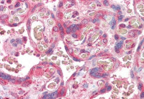
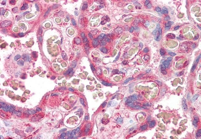
ARG58617 anti-ERV3 antibody IHC-P image
Immunohistochemistry: Paraffin-embedded Human placenta stained with ARG58617 anti-ERV3 antibody at 3.75 µg/ml dilution. Antigen Retrieval: Steam tissue section in Citrate buffer (pH 6.0).