ARG66255
anti-ERK2 antibody [SQab1744]
anti-ERK2 antibody [SQab1744] for Flow cytometry,IHC-Formalin-fixed paraffin-embedded sections,Immunoprecipitation,Western blot and Human,Mouse,Rat
Overview
| Product Description | Recombinant Rabbit Monoclonal antibody [SQab1744] recognizes ERK2 |
|---|---|
| Tested Reactivity | Hu, Ms, Rat |
| Tested Application | FACS, IHC-P, IP, WB |
| Host | Rabbit |
| Clonality | Monoclonal |
| Clone | SQab1744 |
| Isotype | IgG |
| Target Name | ERK2 |
| Antigen Species | Human |
| Immunogen | Synthetic peptide around the C-terminus of Human ERK2. |
| Conjugation | Un-conjugated |
| Alternate Names | MAPK 2; MAPK 1; MAP kinase 2; ERK-2; p41; ERK; MAP kinase 1; PRKM2; PRKM1; EC 2.7.11.24; MAPK2; p40; Extracellular signal-regulated kinase 2; p38; Mitogen-activated protein kinase 2; Mitogen-activated protein kinase 1; ERK2; MAP kinase isoform p42; p42-MAPK; P42MAPK; p41mapk; ERT1 |
Application Instructions
| Application Suggestion |
|
||||||||||
|---|---|---|---|---|---|---|---|---|---|---|---|
| Application Note | IHC-P: Antigen Retrieval: Boil tissue section in Tris/EDTA buffer (pH 9.0). * The dilutions indicate recommended starting dilutions and the optimal dilutions or concentrations should be determined by the scientist. |
||||||||||
| Observed Size | 42 kDa |
Properties
| Form | Liquid |
|---|---|
| Purification | Purification with Protein A. |
| Buffer | PBS, 0.01% Sodium azide, 40% Glycerol and 0.05% BSA. |
| Preservative | 0.01% Sodium azide |
| Stabilizer | 40% Glycerol and 0.05% BSA |
| Storage Instruction | For continuous use, store undiluted antibody at 2-8°C for up to a week. For long-term storage, aliquot and store at -20°C. Storage in frost free freezers is not recommended. Avoid repeated freeze/thaw cycles. Suggest spin the vial prior to opening. The antibody solution should be gently mixed before use. |
| Note | For laboratory research only, not for drug, diagnostic or other use. |
Bioinformation
| Database Links | |
|---|---|
| Gene Symbol | MAPK1 |
| Gene Full Name | mitogen-activated protein kinase 1 |
| Background | ERK2 is a member of the MAP kinase family. MAP kinases, also known as extracellular signal-regulated kinases (ERKs), act as an integration point for multiple biochemical signals, and are involved in a wide variety of cellular processes such as proliferation, differentiation, transcription regulation and development. The activation of this kinase requires its phosphorylation by upstream kinases. Upon activation, this kinase translocates to the nucleus of the stimulated cells, where it phosphorylates nuclear targets. One study also suggests that this protein acts as a transcriptional repressor independent of its kinase activity. The encoded protein has been identified as a moonlighting protein based on its ability to perform mechanistically distinct functions. Two alternatively spliced transcript variants encoding the same protein, but differing in the UTRs, have been reported for this gene. [provided by RefSeq, Jan 2014] |
| Function | Serine/threonine kinase which acts as an essential component of the MAP kinase signal transduction pathway. MAPK1/ERK2 and MAPK3/ERK1 are the 2 MAPKs which play an important role in the MAPK/ERK cascade. They participate also in a signaling cascade initiated by activated KIT and KITLG/SCF. Depending on the cellular context, the MAPK/ERK cascade mediates diverse biological functions such as cell growth, adhesion, survival and differentiation through the regulation of transcription, translation, cytoskeletal rearrangements. The MAPK/ERK cascade plays also a role in initiation and regulation of meiosis, mitosis, and postmitotic functions in differentiated cells by phosphorylating a number of transcription factors. About 160 substrates have already been discovered for ERKs. Many of these substrates are localized in the nucleus, and seem to participate in the regulation of transcription upon stimulation. However, other substrates are found in the cytosol as well as in other cellular organelles, and those are responsible for processes such as translation, mitosis and apoptosis. Moreover, the MAPK/ERK cascade is also involved in the regulation of the endosomal dynamics, including lysosome processing and endosome cycling through the perinuclear recycling compartment (PNRC); as well as in the fragmentation of the Golgi apparatus during mitosis. The substrates include transcription factors (such as ATF2, BCL6, ELK1, ERF, FOS, HSF4 or SPZ1), cytoskeletal elements (such as CANX, CTTN, GJA1, MAP2, MAPT, PXN, SORBS3 or STMN1), regulators of apoptosis (such as BAD, BTG2, CASP9, DAPK1, IER3, MCL1 or PPARG), regulators of translation (such as EIF4EBP1) and a variety of other signaling-related molecules (like ARHGEF2, FRS2 or GRB10). Protein kinases (such as RAF1, RPS6KA1/RSK1, RPS6KA3/RSK2, RPS6KA2/RSK3, RPS6KA6/RSK4, SYK, MKNK1/MNK1, MKNK2/MNK2, RPS6KA5/MSK1, RPS6KA4/MSK2, MAPKAPK3 or MAPKAPK5) and phosphatases (such as DUSP1, DUSP4, DUSP6 or DUSP16) are other substrates which enable the propagation the MAPK/ERK signal to additional cytosolic and nuclear targets, thereby extending the specificity of the cascade. [UniProt] |
| Calculated MW | 41 kDa |
| PTM | Phosphorylated upon KIT and FLT3 signaling (By similarity). Dually phosphorylated on Thr-185 and Tyr-187, which activates the enzyme. Undergoes regulatory phosphorylation on additional residues such as Ser-246 and Ser-248 in the kinase insert domain (KID) These phosphorylations, which are probably mediated by more than one kinase, are important for binding of MAPK1/ERK2 to importin-7 (IPO7) and its nuclear translocation. In addition, autophosphorylation of Thr-190 was shown to affect the subcellular localization of MAPK1/ERK2 as well. Ligand-activated ALK induces tyrosine phosphorylation. Dephosphorylated by PTPRJ at Tyr-187. Phosphorylation on Ser-29 by SGK1 results in its activation by enhancing its interaction with MAP2K1/MEK1 and MAP2K2/MEK2. DUSP3 and DUSP6 dephosphorylate specifically MAPK1/ERK2 and MAPK3/ERK1 whereas DUSP9 dephosphorylates a broader range of MAPKs. ISGylated. [UniProt] |
Images (5) Click the Picture to Zoom In
-
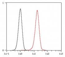
ARG66255 anti-ERK2 antibody [SQab1744] FACS image
Flow Cytometry: HeLa cells were fixed with 4% paraformaldehyde (10 min) and then permeabilized with 0.1% TritonX-100 for 15 min. The cells were stained with ARG66255 anti-ERK2 antibody [SQab1744] (red) at 1:200 in 1x PBS/1% BSA for 30 min at 4°C, followed by Alexa Fluor® 488 labelled secondary antibody. Unlabelled sample (black) was used as a control.
-
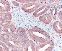
ARG66255 anti-ERK2 antibody [SQab1744] IHC-P image
Immunohistochemistry: Formalin-fixed and paraffin-embedded Endometrium cancer tissue stained with ARG66255 anti-ERK2 antibody [SQab1744] at 1:1600. Antigen Retrieval: Boil tissue section in Tris/EDTA buffer (pH 9.0).
-
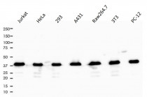
ARG66255 anti-ERK2 antibody [SQab1744] WB image
Western blot: 10 µg of Jurkat, HeLa, 293, A431, Raw264.7, 3T3 and PC-12 cell lysates stained with ARG66255 anti-ERK2 antibody [SQab1744] at 1:1000 dilution.
-
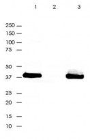
ARG66255 anti-ERK2 antibody [SQab1744] IP image
Immunoprecipitation: ERK2 was immunoprecipitated from 0.4 mg of A431 lysate with ARG66255 anti-ERK2 antibody [SQab1744] at 1:20 dilution.
1. IP by using ARG66255 in A431 whole cell lysate
2. PBS instead of ARG66255 in A431 whole cell lysate
3. 10 µg of A431 whole cell lysate (input) -
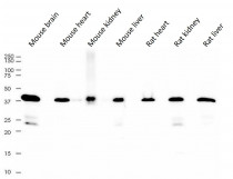
ARG66255 anti-ERK2 antibody [SQab1744] WB image
Western blot: 10 µg of Mouse brain, Mouse heart, Mouse kidney, Mouse liver, Rat heart, Rat kidney and Rat liver lysates stained with ARG66255 anti-ERK2 antibody [SQab1744] at 1:1000 dilution.


