ARG55922
anti-ERBB4 / HER4 phospho (Tyr1162) antibody
anti-ERBB4 / HER4 phospho (Tyr1162) antibody for ICC/IF,IHC-Formalin-fixed paraffin-embedded sections,Western blot and Human
Overview
| Product Description | Rabbit Polyclonal antibody recognizes ERBB4 / HER4 phospho (Tyr1162) |
|---|---|
| Tested Reactivity | Hu |
| Predict Reactivity | Ms, Rat |
| Tested Application | ICC/IF, IHC-P, WB |
| Host | Rabbit |
| Clonality | Polyclonal |
| Isotype | IgG |
| Target Name | ERBB4 / HER4 |
| Antigen Species | Human |
| Immunogen | KLH-conjugated phosphospecific peptide around Tyr1162 of Human ERBB4 / HER4. |
| Conjugation | Un-conjugated |
| Alternate Names | E4ICD; s80HER4; Proto-oncogene-like protein c-ErbB-4; p180erbB4; Tyrosine kinase-type cell surface receptor HER4; Receptor tyrosine-protein kinase erbB-4; 4ICD; EC 2.7.10.1; ALS19; HER4 |
Application Instructions
| Application Suggestion |
|
||||||||
|---|---|---|---|---|---|---|---|---|---|
| Application Note | * The dilutions indicate recommended starting dilutions and the optimal dilutions or concentrations should be determined by the scientist. | ||||||||
| Positive Control | FG pancreatic carcinoma cell + EGF |
Properties
| Form | Liquid |
|---|---|
| Purification | Purification with Protein G and phospho-specific peptide, the non-phospho specific antibodies were removed by chromatography using non-phosphopeptide. |
| Buffer | PBS and 0.09% (W/V) Sodium azide. |
| Preservative | 0.09% (W/V) Sodium azide. |
| Storage Instruction | For continuous use, store undiluted antibody at 2-8°C for up to a week. For long-term storage, aliquot and store at -20°C or below. Storage in frost free freezers is not recommended. Avoid repeated freeze/thaw cycles. Suggest spin the vial prior to opening. The antibody solution should be gently mixed before use. |
| Note | For laboratory research only, not for drug, diagnostic or other use. |
Bioinformation
| Database Links |
Swiss-port # Q15303 Human Receptor tyrosine-protein kinase erbB-4 |
|---|---|
| Gene Symbol | ERBB4 |
| Gene Full Name | erb-b2 receptor tyrosine kinase 4 |
| Background | This gene is a member of the Tyr protein kinase family and the epidermal growth factor receptor subfamily. It encodes a single-pass type I membrane protein with multiple cysteine rich domains, a transmembrane domain, a tyrosine kinase domain, a phosphotidylinositol-3 kinase binding site and a PDZ domain binding motif. The protein binds to and is activated by neuregulins and other factors and induces a variety of cellular responses including mitogenesis and differentiation. Multiple proteolytic events allow for the release of a cytoplasmic fragment and an extracellular fragment. Mutations in this gene have been associated with cancer. Alternatively spliced variants which encode different protein isoforms have been described; however, not all variants have been fully characterized. [provided by RefSeq, Jul 2008] |
| Function | Tyrosine-protein kinase that plays an essential role as cell surface receptor for neuregulins and EGF family members and regulates development of the heart, the central nervous system and the mammary gland, gene transcription, cell proliferation, differentiation, migration and apoptosis. Required for normal cardiac muscle differentiation during embryonic development, and for postnatal cardiomyocyte proliferation. Required for normal development of the embryonic central nervous system, especially for normal neural crest cell migration and normal axon guidance. Required for mammary gland differentiation, induction of milk proteins and lactation. Acts as cell-surface receptor for the neuregulins NRG1, NRG2, NRG3 and NRG4 and the EGF family members BTC, EREG and HBEGF. Ligand binding triggers receptor dimerization and autophosphorylation at specific tyrosine residues that then serve as binding sites for scaffold proteins and effectors. Ligand specificity and signaling is modulated by alternative splicing, proteolytic processing, and by the formation of heterodimers with other ERBB family members, thereby creating multiple combinations of intracellular phosphotyrosines that trigger ligand- and context-specific cellular responses. Mediates phosphorylation of SHC1 and activation of the MAP kinases MAPK1/ERK2 and MAPK3/ERK1. Isoform JM-A CYT-1 and isoform JM-B CYT-1 phosphorylate PIK3R1, leading to the activation of phosphatidylinositol 3-kinase and AKT1 and protect cells against apoptosis. Isoform JM-A CYT-1 and isoform JM-B CYT-1 mediate reorganization of the actin cytoskeleton and promote cell migration in response to NRG1. Isoform JM-A CYT-2 and isoform JM-B CYT-2 lack the phosphotyrosine that mediates interaction with PIK3R1, and hence do not phosphorylate PIK3R1, do not protect cells against apoptosis, and do not promote reorganization of the actin cytoskeleton and cell migration. Proteolytic processing of isoform JM-A CYT-1 and isoform JM-A CYT-2 gives rise to the corresponding soluble intracellular domains (4ICD) that translocate to the nucleus, promote nuclear import of STAT5A, activation of STAT5A, mammary epithelium differentiation, cell proliferation and activation of gene expression. The ERBB4 soluble intracellular domains (4ICD) colocalize with STAT5A at the CSN2 promoter to regulate transcription of milk proteins during lactation. The ERBB4 soluble intracellular domains can also translocate to mitochondria and promote apoptosis. [UniProt] |
| Calculated MW | 147 kDa |
| PTM | Isoform JM-A CYT-1 and isoform JM-A CYT-2 are processed by ADAM17. Proteolytic processing in response to ligand or 12-O-tetradecanoylphorbol-13-acetate stimulation results in the production of 120 kDa soluble receptor forms and intermediate membrane-anchored 80 kDa fragments (m80HER4), which are further processed by a presenilin-dependent gamma-secretase to release a cytoplasmic intracellular domain (E4ICD; E4ICD1/s80Cyt1 or E4ICD2/s80Cyt2, depending on the isoform). Membrane-anchored 80 kDa fragments of the processed isoform JM-A CYT-1 are more readily degraded by the proteasome than fragments of isoform JM-A CYT-2, suggesting a prevalence of E4ICD2 over E4ICD1. Isoform JM-B CYT-1 and isoform JM-B CYT-2 lack the ADAM17 cleavage site and are not processed by ADAM17, precluding further processing by gamma-secretase. Autophosphorylated on tyrosine residues in response to ligand binding. Autophosphorylation occurs in trans, i.e. one subunit of the dimeric receptor phosphorylates tyrosine residues on the other subunit. Ligands trigger phosphorylation at specific tyrosine residues, thereby creating binding sites for scaffold proteins and effectors. Constitutively phosphorylated at a basal level when overexpressed in heterologous systems; ligand binding leads to increased phosphorylation. Phosphorylation at Tyr-1035 is important for interaction with STAT1. Phosphorylation at Tyr-1056 is important for interaction with PIK3R1. Phosphorylation at Tyr-1242 is important for interaction with SHC1. Phosphorylation at Tyr-1188 may also contribute to the interaction with SHC1. Isoform JM-A CYT-2 is constitutively phosphorylated on tyrosine residues in a ligand-independent manner. E4ICD2 but not E4ICD1 is phosphorylated on tyrosine residues. Ubiquitinated. During mitosis, the ERBB4 intracellular domain is ubiquitinated by the APC/C complex and targeted to proteasomal degradation. Isoform JM-A CYT-1 and isoform JM-B CYT-1 are ubiquitinated by WWP1. The ERBB4 intracellular domain (E4ICD1) is ubiquitinated, and this involves NEDD4. |
Images (3) Click the Picture to Zoom In
-
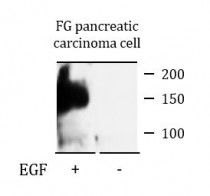
ARG55922 anti-ERBB4 / HER4 phospho (Tyr1162) antibody WB image
Western blot: FG pancreatic carcinoma cell lysate treated or untreated with EGF (50 ng/ml) for 15 min. The blots were stained with ARG55922 anti-ERBB4 / HER4 phospho (Tyr1162) antibody at 1:750 dilution.
-
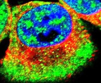
ARG55922 anti-ERBB4 / HER4 phospho (Tyr1162) antibody ICC/IF image
Immunofluorescence: MCF7 cells stained with ARG55922 anti-ERBB4 / HER4 phospho (Tyr1162) antibody (green). Actin filaments have been labeled with Alexa Fluor® 555 conjugated with Phalloidin (red). DAPI (blue) for nuclear staining.
-

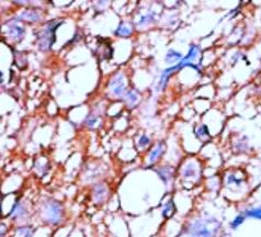
ARG55922 anti-ERBB4 / HER4 phospho (Tyr1162) antibody IHC-P image
Immunohistochemistry: Formalin-fixed and paraffin-embedded Human cancer tissue stained with ARG55922 anti-ERBB4 / HER4 phospho (Tyr1162) antibody.