ARG52859
anti-ERBB4 / HER4 antibody
anti-ERBB4 / HER4 antibody for IHC-Formalin-fixed paraffin-embedded sections,Immunoprecipitation,Western blot and Human,Mouse
Cancer antibody; Controls and Markers antibody; Signaling Transduction antibody
Overview
| Product Description | Rabbit Polyclonal antibody recognizes ERBB4 / HER4 |
|---|---|
| Tested Reactivity | Hu, Ms |
| Tested Application | IHC-P, IP, WB |
| Host | Rabbit |
| Clonality | Polyclonal |
| Isotype | IgG |
| Target Name | ERBB4 / HER4 |
| Antigen Species | Human |
| Immunogen | Synthetic peptide from C-terminus of human c-erbB-4 protein. |
| Conjugation | Un-conjugated |
| Alternate Names | E4ICD; s80HER4; Proto-oncogene-like protein c-ErbB-4; p180erbB4; Tyrosine kinase-type cell surface receptor HER4; Receptor tyrosine-protein kinase erbB-4; 4ICD; EC 2.7.10.1; ALS19; HER4 |
Application Instructions
| Application Suggestion |
|
||||||||
|---|---|---|---|---|---|---|---|---|---|
| Application Note | IHC-P: Incubation Time: 30 min at RT. * The dilutions indicate recommended starting dilutions and the optimal dilutions or concentrations should be determined by the scientist. |
||||||||
| Positive Control | Breast Carcinoma, A431 |
Properties
| Form | Liquid |
|---|---|
| Purification | Immunogen affinity purified |
| Buffer | PBS (pH 7.6), 1% BSA and < 0.1% Sodium azide |
| Preservative | < 0.1% Sodium azide |
| Stabilizer | 1% BSA |
| Storage Instruction | For continuous use, store undiluted antibody at 2-8°C for up to a week. For long-term storage, aliquot and store at -20°C or below. Storage in frost free freezers is not recommended. Avoid repeated freeze/thaw cycles. Suggest spin the vial prior to opening. The antibody solution should be gently mixed before use. |
| Note | For laboratory research only, not for drug, diagnostic or other use. |
Bioinformation
| Database Links |
Swiss-port # Q15303 Human Receptor tyrosine-protein kinase erbB-4 Swiss-port # Q61527 Mouse Receptor tyrosine-protein kinase erbB-4 |
|---|---|
| Background | c-erbB-4 is the fourth member of class 1 receptor kinase family. HER-4 is most predominantly expressed in several breast carcinoma cell lines, and in normal skeletal muscle, heart, pituitary, brain, and cerebellum. Breast tumor cell lines T47-D, MDA-MB-453, BT-474 and H3396 are found to have the highest levels of mRNA, and intermediate levels are seen in MCF-7, MDA-MB-330 and MDA-MB-361. Expression of erbB-4 is low or absent in some breast tumor cell lines such as MDA-MB-231, MDA-MB-157, MDA-MB-468, and SKBR-3. |
| Cellular Localization | Cytoplasm, Membrane |
| Research Area | Cancer antibody; Controls and Markers antibody; Signaling Transduction antibody |
| Calculated MW | 147 kDa |
| PTM | Isoform JM-A CYT-1 and isoform JM-A CYT-2 are processed by ADAM17. Proteolytic processing in response to ligand or 12-O-tetradecanoylphorbol-13-acetate stimulation results in the production of 120 kDa soluble receptor forms and intermediate membrane-anchored 80 kDa fragments (m80HER4), which are further processed by a presenilin-dependent gamma-secretase to release a cytoplasmic intracellular domain (E4ICD; E4ICD1/s80Cyt1 or E4ICD2/s80Cyt2, depending on the isoform). Membrane-anchored 80 kDa fragments of the processed isoform JM-A CYT-1 are more readily degraded by the proteasome than fragments of isoform JM-A CYT-2, suggesting a prevalence of E4ICD2 over E4ICD1. Isoform JM-B CYT-1 and isoform JM-B CYT-2 lack the ADAM17 cleavage site and are not processed by ADAM17, precluding further processing by gamma-secretase. Autophosphorylated on tyrosine residues in response to ligand binding. Autophosphorylation occurs in trans, i.e. one subunit of the dimeric receptor phosphorylates tyrosine residues on the other subunit. Ligands trigger phosphorylation at specific tyrosine residues, thereby creating binding sites for scaffold proteins and effectors. Constitutively phosphorylated at a basal level when overexpressed in heterologous systems; ligand binding leads to increased phosphorylation. Phosphorylation at Tyr-1035 is important for interaction with STAT1. Phosphorylation at Tyr-1056 is important for interaction with PIK3R1. Phosphorylation at Tyr-1242 is important for interaction with SHC1. Phosphorylation at Tyr-1188 may also contribute to the interaction with SHC1. Isoform JM-A CYT-2 is constitutively phosphorylated on tyrosine residues in a ligand-independent manner. E4ICD2 but not E4ICD1 is phosphorylated on tyrosine residues. Ubiquitinated. During mitosis, the ERBB4 intracellular domain is ubiquitinated by the APC/C complex and targeted to proteasomal degradation. Isoform JM-A CYT-1 and isoform JM-B CYT-1 are ubiquitinated by WWP1. The ERBB4 intracellular domain (E4ICD1) is ubiquitinated, and this involves NEDD4. |
Images (2) Click the Picture to Zoom In
-

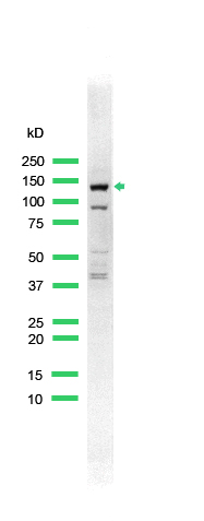
ARG52859 anti-ERBB4 / HER4 antibody WB image
Western blot: A431 cell lysate stained with ARG52859 anti-ERBB4 / HER4 antibody.
-
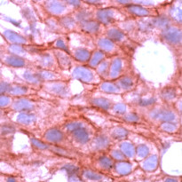
ARG52859 anti-ERBB4 / HER4 antibody IHC-P image
Immunohistochemistry: Human Breast Carcinoma stained with ARG52859 anti-ERBB4 / HER4 antibody.

Specific References
HER4 promotes the progression of colorectal cancer by promoting epithelial‑mesenchymal transition.
IHC-P / Human


