ARG55546
anti-ERBB3 / HER3 antibody
anti-ERBB3 / HER3 antibody for Flow cytometry,IHC-Formalin-fixed paraffin-embedded sections,Western blot and Human,Mouse
Cancer antibody; Controls and Markers antibody; Signaling Transduction antibody
Overview
| Product Description | Rabbit Polyclonal antibody recognizes ERBB3 / HER3 |
|---|---|
| Tested Reactivity | Hu, Ms |
| Tested Application | FACS, IHC-P, WB |
| Host | Rabbit |
| Clonality | Polyclonal |
| Isotype | IgG |
| Target Name | ERBB3 / HER3 |
| Antigen Species | Human |
| Immunogen | KLH-conjugated synthetic peptide corresponding to aa. 24-55 (N-terminus) of Human ERBB3. |
| Conjugation | Un-conjugated |
| Alternate Names | MDA-BF-1; LCCS2; p180-ErbB3; Proto-oncogene-like protein c-ErbB-3; c-erbB3; p85-sErbB3; Tyrosine kinase-type cell surface receptor HER3; p45-sErbB3; erbB3-S; HER3; c-erbB-3; ErbB-3; EC 2.7.10.1; Receptor tyrosine-protein kinase erbB-3 |
Application Instructions
| Application Suggestion |
|
||||||||
|---|---|---|---|---|---|---|---|---|---|
| Application Note | * The dilutions indicate recommended starting dilutions and the optimal dilutions or concentrations should be determined by the scientist. | ||||||||
| Positive Control | Mouse brain |
Properties
| Form | Liquid |
|---|---|
| Purification | Purification with Protein G. |
| Buffer | PBS and 0.09% (W/V) Sodium azide |
| Preservative | 0.09% (W/V) Sodium azide |
| Storage Instruction | For continuous use, store undiluted antibody at 2-8°C for up to a week. For long-term storage, aliquot and store at -20°C or below. Storage in frost free freezers is not recommended. Avoid repeated freeze/thaw cycles. Suggest spin the vial prior to opening. The antibody solution should be gently mixed before use. |
| Note | For laboratory research only, not for drug, diagnostic or other use. |
Bioinformation
| Database Links |
Swiss-port # P21860 Human Receptor tyrosine-protein kinase erbB-3 Swiss-port # Q61526 Mouse Receptor tyrosine-protein kinase erbB-3 |
|---|---|
| Gene Symbol | ERBB3 |
| Gene Full Name | erb-b2 receptor tyrosine kinase 3 |
| Background | This gene encodes a member of the epidermal growth factor receptor (EGFR) family of receptor tyrosine kinases. This membrane-bound protein has a neuregulin binding domain but not an active kinase domain. It therefore can bind this ligand but not convey the signal into the cell through protein phosphorylation. However, it does form heterodimers with other EGF receptor family members which do have kinase activity. Heterodimerization leads to the activation of pathways which lead to cell proliferation or differentiation. Amplification of this gene and/or overexpression of its protein have been reported in numerous cancers, including prostate, bladder, and breast tumors. Alternate transcriptional splice variants encoding different isoforms have been characterized. One isoform lacks the intermembrane region and is secreted outside the cell. This form acts to modulate the activity of the membrane-bound form. Additional splice variants have also been reported, but they have not been thoroughly characterized. [provided by RefSeq, Jul 2008] |
| Function | Binds and is activated by neuregulins and NTAK. May also be activated by CSPG5. [UniProt] |
| Cellular Localization | Isoform 1: Cell membrane; Single-pass type I membrane protein |
| Research Area | Cancer antibody; Controls and Markers antibody; Signaling Transduction antibody |
| Calculated MW | 148 kDa |
| PTM | Autophosphorylated (PubMed:20351256). Ligand-binding increases phosphorylation on tyrosine residues and promotes its association with the p85 subunit of phosphatidylinositol 3-kinase (PubMed:20682778). |
Images (3) Click the Picture to Zoom In
-
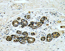
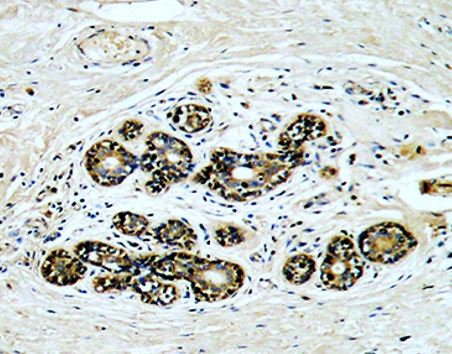
ARG55546 anti-ERBB3 / HER3 antibody IHC-P image
Immunohistochemistry: Formalin-fixed and paraffin-embedded Human breast carcinoma stained with ARG55546 anti-ERBB3 / HER3 antibody.
-

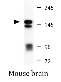
ARG55546 anti-ERBB3 / HER3 antibody WB image
Western blot: Mouse brain lysate stained with ARG55546 anti-ERBB3 / HER3 antibody.
-
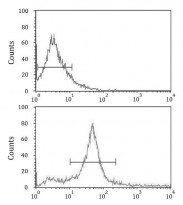
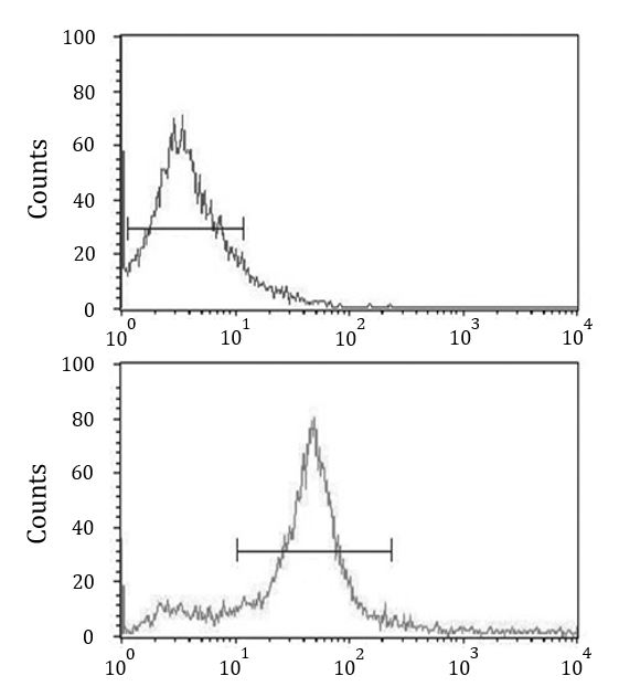
ARG55546 anti-ERBB3 / HER3 antibody FACS image
Flow Cytometry: 293 cells stained with ARG55546 anti-ERBB3 / HER3 antibody (bottom histogram) or without primary antibody control (top histogram), followed by incubation with FITC labelled secondary antibody.