ARG66498
anti-ERBB2 / HER2 antibody
anti-ERBB2 / HER2 antibody for IHC-Formalin-fixed paraffin-embedded sections and Human
Cancer antibody; Controls and Markers antibody; Signaling Transduction antibody; Circulating Tumor Cells BioMarker antibody
Overview
| Product Description | Mouse Monoclonal antibody recognizes ERBB2 / HER2 |
|---|---|
| Tested Reactivity | Hu |
| Tested Application | IHC-P |
| Host | Mouse |
| Clonality | Monoclonal |
| Isotype | IgG2b, kappa |
| Target Name | ERBB2 / HER2 |
| Antigen Species | Human |
| Immunogen | Synthetic peptide derived from Human ERBB2 / HER2. |
| Conjugation | Un-conjugated |
| Alternate Names | p185erbB2; Proto-oncogene c-ErbB-2; Metastatic lymph node gene 19 protein; Proto-oncogene Neu; NGL; EC 2.7.10.1; CD340; CD antigen CD340; TKR1; HER-2; Tyrosine kinase-type cell surface receptor HER2; HER2; NEU; HER-2/neu; MLN 19; Receptor tyrosine-protein kinase erbB-2 |
Application Instructions
| Application Suggestion |
|
||||
|---|---|---|---|---|---|
| Application Note | IHC-P: Antigen Retrieval: Tris/EDTA buffer (pH 8.0) was used. * The dilutions indicate recommended starting dilutions and the optimal dilutions or concentrations should be determined by the scientist. |
Properties
| Form | Liquid |
|---|---|
| Purification | Affinity purification with immunogen. |
| Buffer | PBS, 0.02% Sodium azide, 50% Glycerol and 0.5% BSA. |
| Preservative | 0.02% Sodium azide |
| Stabilizer | 50% Glycerol and 0.5% BSA |
| Concentration | 1 mg/ml |
| Storage Instruction | For continuous use, store undiluted antibody at 2-8°C for up to a week. For long-term storage, aliquot and store at -20°C. Storage in frost free freezers is not recommended. Avoid repeated freeze/thaw cycles. Suggest spin the vial prior to opening. The antibody solution should be gently mixed before use. |
| Note | For laboratory research only, not for drug, diagnostic or other use. |
Bioinformation
| Database Links |
Swiss-port # P04626 Human Receptor tyrosine-protein kinase erbB-2 |
|---|---|
| Gene Symbol | ERBB2 |
| Gene Full Name | erb-b2 receptor tyrosine kinase 2 |
| Background | This gene encodes a member of the epidermal growth factor (EGF) receptor family of receptor tyrosine kinases. This protein has no ligand binding domain of its own and therefore cannot bind growth factors. However, it does bind tightly to other ligand-bound EGF receptor family members to form a heterodimer, stabilizing ligand binding and enhancing kinase-mediated activation of downstream signalling pathways, such as those involving mitogen-activated protein kinase and phosphatidylinositol-3 kinase. Allelic variations at amino acid positions 654 and 655 of isoform a (positions 624 and 625 of isoform b) have been reported, with the most common allele, Ile654/Ile655, shown here. Amplification and/or overexpression of this gene has been reported in numerous cancers, including breast and ovarian tumors. Alternative splicing results in several additional transcript variants, some encoding different isoforms and others that have not been fully characterized. [provided by RefSeq, Jul 2008] |
| Function | Protein tyrosine kinase that is part of several cell surface receptor complexes, but that apparently needs a coreceptor for ligand binding. Essential component of a neuregulin-receptor complex, although neuregulins do not interact with it alone. GP30 is a potential ligand for this receptor. Regulates outgrowth and stabilization of peripheral microtubules (MTs). Upon ERBB2 activation, the MEMO1-RHOA-DIAPH1 signaling pathway elicits the phosphorylation and thus the inhibition of GSK3B at cell membrane. This prevents the phosphorylation of APC and CLASP2, allowing its association with the cell membrane. In turn, membrane-bound APC allows the localization of MACF1 to the cell membrane, which is required for microtubule capture and stabilization. In the nucleus is involved in transcriptional regulation. Associates with the 5'-TCAAATTC-3' sequence in the PTGS2/COX-2 promoter and activates its transcription. Implicated in transcriptional activation of CDKN1A; the function involves STAT3 and SRC. Involved in the transcription of rRNA genes by RNA Pol I and enhances protein synthesis and cell growth. [UniProt] |
| Cellular Localization | Isoform 1: Cell membrane; Single-pass type I membrane protein. Cytoplasm, perinuclear region. Nucleus. Note=Translocation to the nucleus requires endocytosis, probably endosomal sorting and is mediated by importin beta-1/KPNB1. Isoform 2: Cytoplasm. Nucleus. Isoform 3: Cytoplasm. Nucleus. [UniProt] |
| Research Area | Cancer antibody; Controls and Markers antibody; Signaling Transduction antibody; Circulating Tumor Cells BioMarker antibody |
| Calculated MW | 138 kDa |
| PTM | Autophosphorylated. Autophosphorylation occurs in trans, i.e. one subunit of the dimeric receptor phosphorylates tyrosine residues on the other subunit (Probable). Ligand-binding increases phosphorylation on tyrosine residues (PubMed:27134172). Signaling via SEMA4C promotes phosphorylation at Tyr-1248 (PubMed:17554007). Dephosphorylated by PTPN12 (PubMed:27134172). [UniProt] |
Images (1) Click the Picture to Zoom In
-
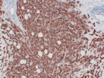
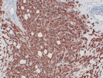
ARG66498 anti-ERBB2 / HER2 antibody IHC-P image
Immunohistochemistry: Paraffin-embedded Human breast carcinoma stained with ARG66498 anti-ERBB2 / HER2 antibody at 1:200 (4°C, overnight). Antigen Retrieval: Tris/EDTA buffer (pH 8.0) was used.