ARG58024
anti-ERAB / HADH2 antibody
anti-ERAB / HADH2 antibody for Flow cytometry,ICC/IF,IHC-Formalin-fixed paraffin-embedded sections,Western blot and Human,Mouse
Overview
| Product Description | Rabbit Polyclonal antibody recognizes ERAB / HADH2 |
|---|---|
| Tested Reactivity | Hu, Ms |
| Tested Application | FACS, ICC/IF, IHC-P, WB |
| Host | Rabbit |
| Clonality | Polyclonal |
| Isotype | IgG |
| Target Name | ERAB / HADH2 |
| Antigen Species | Human |
| Immunogen | Partial recombinant protein corresponding to aa. 48-261 of Human ERAB / HADH2. |
| Conjugation | Un-conjugated |
| Alternate Names | MRPP2; ERAB; ABAD; Short chain dehydrogenase/reductase family 5C member 1; HCD2; SDR5C1; 3-hydroxyacyl-CoA dehydrogenase type-2; 17-beta-hydroxysteroid dehydrogenase 10; SCHAD; 3-hydroxy-2-methylbutyryl-CoA dehydrogenase; EC 1.1.1.178; Short-chain type dehydrogenase/reductase XH98G2; Endoplasmic reticulum-associated amyloid beta-peptide-binding protein; 17-beta-HSD 10; CAMR; Mitochondrial RNase P protein 2; EC 1.1.1.35; Type II HADH; 3-hydroxyacyl-CoA dehydrogenase type II; EC 1.1.1.51; 17b-HSD10; MRX31; DUPXp11.22; HADH2; MRX17; MRXS10; Mitochondrial ribonuclease P protein 2; MHBD |
Application Instructions
| Application Suggestion |
|
||||||||||
|---|---|---|---|---|---|---|---|---|---|---|---|
| Application Note | IHC-P: Antigen Retrieval: Steam tissue section in Citrate buffer (pH 6.0) for 20 min followed by cooling at RT. * The dilutions indicate recommended starting dilutions and the optimal dilutions or concentrations should be determined by the scientist. |
Properties
| Form | Liquid |
|---|---|
| Purification | Affinity purification with immunogen. |
| Buffer | PBS, 0.025% Sodium azide and 2.5% BSA. |
| Preservative | 0.025% Sodium azide |
| Stabilizer | 2.5% BSA |
| Concentration | 0.5 mg/ml |
| Storage Instruction | For continuous use, store undiluted antibody at 2-8°C for up to a week. For long-term storage, aliquot and store at -20°C or below. Storage in frost free freezers is not recommended. Avoid repeated freeze/thaw cycles. Suggest spin the vial prior to opening. The antibody solution should be gently mixed before use. |
| Note | For laboratory research only, not for drug, diagnostic or other use. |
Bioinformation
| Database Links |
Swiss-port # Q99714 Human 3-hydroxyacyl-CoA dehydrogenase type-2 |
|---|---|
| Gene Symbol | HSD17B10 |
| Gene Full Name | hydroxysteroid (17-beta) dehydrogenase 10 |
| Background | This gene encodes 3-hydroxyacyl-CoA dehydrogenase type II, a member of the short-chain dehydrogenase/reductase superfamily. The gene product is a mitochondrial protein that catalyzes the oxidation of a wide variety of fatty acids and steroids, and is a subunit of mitochondrial ribonuclease P, which is involved in tRNA maturation. The protein has been implicated in the development of Alzheimer disease, and mutations in the gene are the cause of 17beta-hydroxysteroid dehydrogenase type 10 (HSD10) deficiency. Several alternatively spliced transcript variants have been identified, but the full-length nature of only two transcript variants has been determined. [provided by RefSeq, Aug 2014] |
| Function | Functions in mitochondrial tRNA maturation. Part of mitochondrial ribonuclease P, an enzyme composed of MRPP1/TRMT10C, MRPP2/HSD17B10 and MRPP3/KIAA0391, which cleaves tRNA molecules in their 5'-ends. Catalyzes the beta-oxidation at position 17 of androgens and estrogens and has 3-alpha-hydroxysteroid dehydrogenase activity with androsterone. Catalyzes the third step in the beta-oxidation of fatty acids. Carries out oxidative conversions of 7-alpha-OH and 7-beta-OH bile acids. Also exhibits 20-beta-OH and 21-OH dehydrogenase activities with C21 steroids. By interacting with intracellular amyloid-beta, it may contribute to the neuronal dysfunction associated with Alzheimer disease (AD). [UniProt] |
| Calculated MW | 27 kDa |
Images (9) Click the Picture to Zoom In
-
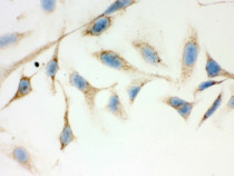
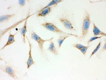
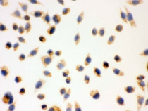
ARG58024 anti-ERAB / HADH2 antibody ICC/IF image
Immunocytochemistry: HeLa cells stained with ARG58024 anti-ERAB / HADH2 antibody.
-
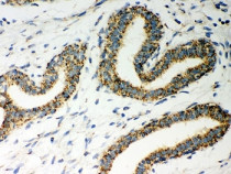
ARG58024 anti-ERAB / HADH2 antibody IHC-P image
Immunohistochemistry: Paraffin-embedded Human breast cancer tissue stained with ARG58024 anti-ERAB / HADH2 antibody. Antigen Retrieval: Steam tissue section in Citrate buffer (pH 6.0) for 20 min followed by cooling at RT.
-
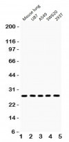
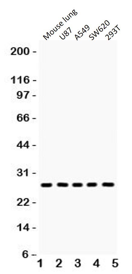
ARG58024 anti-ERAB / HADH2 antibody WB image
Western blot: 1) Mouse lung, 2) U87, 3) A549, 4) SW620 and 5) 293T cell lysates stained with ARG58024 anti-ERAB / HADH2 antibody.
-

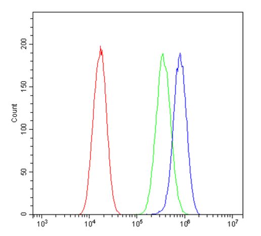
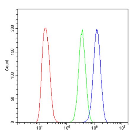
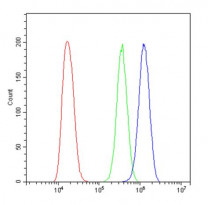
ARG58024 anti-ERAB / HADH2 antibody FACS image
Flow Cytometry: A549 cells were blocked with 10% normal goat serum and then stained with ARG58024 anti-ERAB / HADH2 antibody (blue) at 1 μg/10^6 cells for 30 min at 20°C, followed by incubation with Alexa Fluor® 488 labelled secondary antibody. Isotype control antibody (green) was Rabbit IgG (1 μg/10^6) used under the same conditions. Unlabelled sample (red) was also used as a control.
-
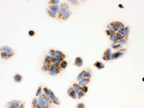
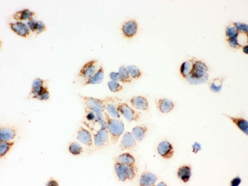
ARG58024 anti-ERAB / HADH2 antibody ICC/IF image
Immunocytochemistry: MCF7 cells stained with ARG58024 anti-ERAB / HADH2 antibody.
-

ARG58024 anti-ERAB / HADH2 antibody ICC/IF image
Immunocytochemistry: Human MCF7 cells stained with ARG58024 anti-ERAB / HADH2 antibody.
-

ARG58024 anti-ERAB / HADH2 antibody ICC/IF image
Immunocytochemistry: Human MCF7 cells stained with ARG58024 anti-ERAB / HADH2 antibody.
-
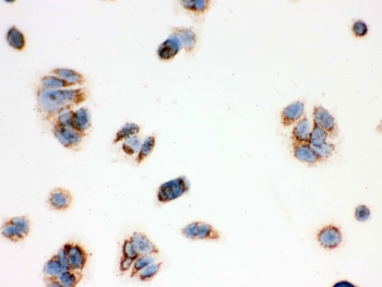
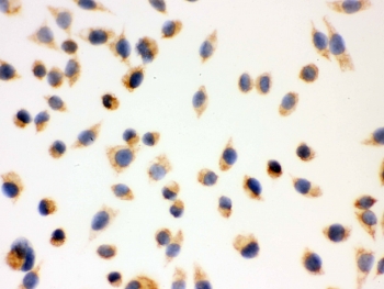
ARG58024 anti-ERAB / HADH2 antibody ICC/IF image
Immunocytochemistry: Human SMMC-7721 cells stained with ARG58024 anti-ERAB / HADH2 antibody.
-
ARG58024 anti-ERAB / HADH2 antibody FACS image
Flow Cytometry: A431 cells were blocked with 10% normal goat serum and then stained with ARG58024 anti-ERAB / HADH2 antibody (blue) at 1 μg/10^6 cells for 30 min at 20°C, followed by incubation with Alexa Fluor® 488 labelled secondary antibody. Isotype control antibody (green) was Rabbit IgG (1 μg/10^6) used under the same conditions. Unlabelled sample (red) was also used as a control.