ARG55576
anti-EPO Receptor antibody
anti-EPO Receptor antibody for Flow cytometry,Western blot and Human
Cell Biology and Cellular Response antibody; Cell Death antibody; Developmental Biology antibody; Immune System antibody; Signaling Transduction antibody
Overview
| Product Description | Rabbit Polyclonal antibody recognizes EPO Receptor |
|---|---|
| Tested Reactivity | Hu |
| Tested Application | FACS, WB |
| Host | Rabbit |
| Clonality | Polyclonal |
| Isotype | IgG |
| Target Name | EPO Receptor |
| Antigen Species | Human |
| Immunogen | KLH-conjugated synthetic peptide corresponding to aa. 470-504 (C-terminus) of Human EPO Receptor. |
| Conjugation | Un-conjugated |
| Alternate Names | Erythropoietin receptor; EPO-R |
Application Instructions
| Application Suggestion |
|
||||||
|---|---|---|---|---|---|---|---|
| Application Note | * The dilutions indicate recommended starting dilutions and the optimal dilutions or concentrations should be determined by the scientist. | ||||||
| Positive Control | K562 |
Properties
| Form | Liquid |
|---|---|
| Purification | Purification with Protein A and immunogen peptide. |
| Buffer | PBS and 0.09% (W/V) Sodium azide |
| Preservative | 0.09% (W/V) Sodium azide |
| Storage Instruction | For continuous use, store undiluted antibody at 2-8°C for up to a week. For long-term storage, aliquot and store at -20°C or below. Storage in frost free freezers is not recommended. Avoid repeated freeze/thaw cycles. Suggest spin the vial prior to opening. The antibody solution should be gently mixed before use. |
| Note | For laboratory research only, not for drug, diagnostic or other use. |
Bioinformation
| Database Links | |
|---|---|
| Gene Symbol | EPOR |
| Gene Full Name | erythropoietin receptor |
| Background | This gene encodes the erythropoietin receptor which is a member of the cytokine receptor family. Upon erythropoietin binding, this receptor activates Jak2 tyrosine kinase which activates different intracellular pathways including: Ras/MAP kinase, phosphatidylinositol 3-kinase and STAT transcription factors. The stimulated erythropoietin receptor appears to have a role in erythroid cell survival. Defects in the erythropoietin receptor may produce erythroleukemia and familial erythrocytosis. Dysregulation of this gene may affect the growth of certain tumors. Alternate splicing results in multiple transcript variants.[provided by RefSeq, May 2010] |
| Function | Receptor for erythropoietin. Mediates erythropoietin-induced erythroblast proliferation and differentiation. Upon EPO stimulation, EPOR dimerizes triggering the JAK2/STAT5 signaling cascade. In some cell types, can also activate STAT1 and STAT3. May also activate the LYN tyrosine kinase. Isoform EPOR-T acts as a dominant-negative receptor of EPOR-mediated signaling. [UniProt] |
| Cellular Localization | Cell membrane; Single-pass type I membrane protein |
| Research Area | Cell Biology and Cellular Response antibody; Cell Death antibody; Developmental Biology antibody; Immune System antibody; Signaling Transduction antibody |
| Calculated MW | 55 kDa |
| PTM | On EPO stimulation, phosphorylated on C-terminal tyrosine residues by JAK2. The phosphotyrosine motifs are also recruitment sites for several SH2-containing proteins and adapter proteins which mediate cell proliferation. Phosphorylation on Tyr-454 is required for PTPN6 interaction, Tyr-426 for PTPN11. Tyr-426 is also required for SOCS3 binding, but Tyr-454/Tyr-456 motif is the preferred binding site. Ubiquitination at Lys-281 mediates receptor internalization, whereas ubiquitination at Lys-453 promotes trafficking of activated receptors to the lysosomes for degradation (By similarity). Ubiquitinated by NOSIP; appears to be either multi-monoubiquitinated or polyubiquitinated. Ubiquitination mediates proliferation and survival of EPO-dependent cells. |
Images (2) Click the Picture to Zoom In
-
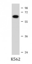
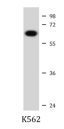
ARG55576 anti-EPO Receptor antibody WB image
Western blot: 20 µg of K562 cell lysate stained with ARG55576 anti-EPO Receptor antibody at 1:1000 dilution.
-

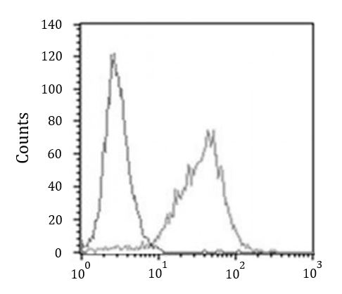
ARG55576 anti-EPO Receptor antibody FACS image
Flow Cytometry: K562 cells stained with ARG55576 anti-EPO Receptor antibody (right histogram) at 1:25 dilution or isotype control antibody (left histogram), followed by incubation with Alexa Fluor® 488 labelled secondary antibody.