ARG22476
anti-EMR1 F4 / 80 antibody [Cl:A3-1]
anti-EMR1 F4 / 80 antibody [Cl:A3-1] for Electron microscopy,Flow cytometry,ICC/IF,IHC-Frozen sections,IHC-Formalin-fixed paraffin-embedded sections,IHC-Resin sections,Immunoprecipitation,Radioimmunoassay,Western blot and Mouse,Rat
Mouse Inflammatory Cell Marker antibody; Macrophage Marker antibody
Overview
| Product Description | Rat Monoclonal antibody [Cl:A3-1] recognizes EMR1 F4 / 80 |
|---|---|
| Tested Reactivity | Ms, Rat |
| Tested Application | EM, FACS, ICC/IF, IHC-Fr, IHC-P, IHC-R, IP, RIA, WB |
| Specificity | EMR1 F4/80 antibody, clone Cl:A3-1, is the best macrophage and microglial marker. It recognises the murine F4/80 antigen, a 160 kD cell surface glycoprotein, member of the EGF-TM7 family. |
| Host | Rat |
| Clonality | Monoclonal |
| Clone | Cl:A3-1 |
| Isotype | IgG2b |
| Target Name | EMR1 F4 / 80 |
| Antigen Species | Mouse |
| Immunogen | Thioglycollate stimulated peritoneal macrophages from C57BL/6 mice. |
| Conjugation | Un-conjugated |
| Alternate Names | EGF-like module-containing mucin-like hormone receptor-like 1; EMR1 hormone receptor; EMR1; Adhesion G protein-coupled receptor E1; EGF-like module receptor 1; TM7LN3 |
Application Instructions
| Application Suggestion |
|
||||||||||||||||||||
|---|---|---|---|---|---|---|---|---|---|---|---|---|---|---|---|---|---|---|---|---|---|
| Application Note | IHC-P: Antigen Retrieval: Proteinase K is recommended for tissues fixed for less than 24 hours. Citrate buffer pH 6.0 is recommended for tissues fixed for more than 24 hours. * The dilutions indicate recommended starting dilutions and the optimal dilutions or concentrations should be determined by the scientist. |
Properties
| Form | Liquid |
|---|---|
| Purification | Purification with Protein G. |
| Buffer | PBS and 0.09% Sodium azide. |
| Preservative | 0.09% Sodium azide |
| Concentration | 1 mg/ml |
| Storage Instruction | For continuous use, store undiluted antibody at 2-8°C for up to a week. For long-term storage, aliquot and store at -20°C or below. Storage in frost free freezers is not recommended. Avoid repeated freeze/thaw cycles. Suggest spin the vial prior to opening. The antibody solution should be gently mixed before use. |
| Note | For laboratory research only, not for drug, diagnostic or other use. |
Bioinformation
| Database Links |
Swiss-port # Q5Y4N8 Rat Adhesion G protein-coupled receptor E2 Swiss-port # Q61549 Mouse Adhesion G protein-coupled receptor E1 |
|---|---|
| Gene Symbol | Adgre1 |
| Gene Full Name | adhesion G protein-coupled receptor E1 |
| Background | EMR1 F4/80 has a domain resembling seven transmembrane G protein-coupled hormone receptors (7TM receptors) at its C-terminus. The N-terminus of the encoded protein has six EGF-like modules, separated from the transmembrane segments by a serine/threonine-rich domain, a feature reminiscent of mucin-like, single-span, integral membrane glycoproteins with adhesive properties. Multiple alternatively spliced transcript variants encoding different isoforms have been found for this gene. [provided by RefSeq, Jan 2012] |
| Function | EMR1 F4/80: Orphan receptor involved in cell adhesion and probably in cell-cell interactions specifically involving cells of the immune system. May play a role in regulatory T-cells (Treg) development. [UniProt] |
| Highlight | Related Antibody Duos and Panels: ARG30326 Inflammatory Cell Antibody Panel (for mouse) Related products: EMR1 F4/80 antibodies; EMR1 F4/80 Duos / Panels; Anti-Rat IgG secondary antibodies; Related news: Exploring Antiviral Immune Response |
| Research Area | Mouse Inflammatory Cell Marker antibody; Macrophage Marker antibody |
| Calculated MW | 98 kDa |
Images (4) Click the Picture to Zoom In
-
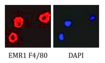
ARG22476 anti-EMR1 F4/80 antibody [Cl:A3-1] ICC/IF image
Immunofluorescence: Rat tumor macrophages stained with ARG22476 anti-EMR1 F4/80 antibody [Cl:A3-1].
-
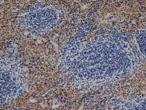
ARG22476 anti-EMR1 F4/80 antibody [Cl:A3-1] IHC-P image
Immunohistochemistry: Mouse spleen cryosection stained with ARG22476 anti-EMR1 F4/80 antibody [Cl:A3-1] at 1:100 dilution.
-
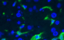
ARG22476 anti-EMR1 F4/80 antibody [Cl:A3-1] IHC-Fr image
Immunohistochemistry: Frozen section of Mouse liver tissue stained with ARG22476 anti-EMR1 F4/80 antibody [Cl:A3-1] at 1:100 dilution.
-
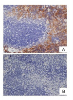
ARG22476 anti-EMR1 F4/80 antibody [Cl:A3-1] IHC-P image
Immunohistochemistry: A. Mouse spleen stained with ARG22476 anti-EMR1 F4/80 antibody [Cl:A3-1] at 1:100 dilution. B. Primary antibodies preincubated with 2 molar excess of Human anti Idiotypic.
Specific References


