ARG63661
anti-EHD2 antibody
anti-EHD2 antibody for Flow cytometry,ICC/IF,IHC-Formalin-fixed paraffin-embedded sections,Western blot and Human
Gene Regulation antibody
Overview
| Product Description | Goat Polyclonal antibody recognizes EHD2 |
|---|---|
| Tested Reactivity | Hu |
| Predict Reactivity | Ms, Rat, Cow, Dog |
| Tested Application | FACS, ICC/IF, IHC-P, WB |
| Specificity | This antibody is expected to recognise EHD1 protein as well as EHD2. |
| Host | Goat |
| Clonality | Polyclonal |
| Isotype | IgG |
| Target Name | EHD2 |
| Antigen Species | Human |
| Immunogen | CRLVPPSKRRHKGSA |
| Conjugation | Un-conjugated |
| Alternate Names | PAST2; EH domain-containing protein 2; PAST homolog 2 |
Application Instructions
| Application Suggestion |
|
||||||||||
|---|---|---|---|---|---|---|---|---|---|---|---|
| Application Note | WB: Recommend incubate at RT for 1h. IHC-P: Antigen Retrieval: Steam tissue section in Citrate buffer (pH 6.0). * The dilutions indicate recommended starting dilutions and the optimal dilutions or concentrations should be determined by the scientist. |
Properties
| Form | Liquid |
|---|---|
| Purification | Purified from goat serum by ammonium sulphate precipitation followed by antigen affinity chromatography using the immunizing peptide. |
| Buffer | Tris saline (pH 7.3), 0.02% Sodium azide and 0.5% BSA |
| Preservative | 0.02% Sodium azide |
| Stabilizer | 0.5% BSA |
| Concentration | 0.5 mg/ml |
| Storage Instruction | For continuous use, store undiluted antibody at 2-8°C for up to a week. For long-term storage, aliquot and store at -20°C or below. Storage in frost free freezers is not recommended. Avoid repeated freeze/thaw cycles. Suggest spin the vial prior to opening. The antibody solution should be gently mixed before use. |
| Note | For laboratory research only, not for drug, diagnostic or other use. |
Bioinformation
| Database Links | |
|---|---|
| Background | This gene encodes a member of the EH domain-containing protein family. These proteins are characterized by a C-terminal EF-hand domain, a nucleotide-binding consensus site at the N terminus and a bipartite nuclear localization signal. The encoded protein interacts with the actin cytoskeleton through an N-terminal domain and also binds to an EH domain-binding protein through the C-terminal EH domain. This interaction appears to connect clathrin-dependent endocytosis to actin, suggesting that this gene product participates in the endocytic pathway. [provided by RefSeq, Jul 2008] |
| Research Area | Gene Regulation antibody |
| Calculated MW | 61 kDa |
Images (8) Click the Picture to Zoom In
-

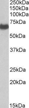
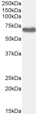
ARG63661 anti-EHD2 antibody WB image
Western Blot: Human Placenta lysate (35 µg protein in RIPA buffer) stained with ARG63661 anti-EHD2 antibody at 0.1 µg/ml dilution.
-
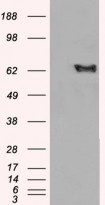
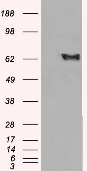
ARG63661 anti-EHD2 antibody WB image
Western Blot: 1). Mock transfection; 2) EHD2 (RC204848) expressing plasmid transfected HEK293 cell lysate standed with ARG63661 anti-EHD2 antibody
-

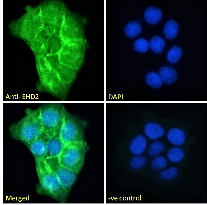
ARG63661 anti-EHD2 antibody ICC/IF image
Immunofluorescence: Paraformaldehyde fixed A431 cells permeabilized with 0.15% Triton. Cells were stained with ARG63661 anti-EHD2 antibody (green) at 10 µg/ml dilution for 1 hour. DAPI (blue) for nuclear staining. Negative control: Unimmunized goat IgG (green) at 10 µg/ml dilution.
-
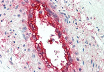
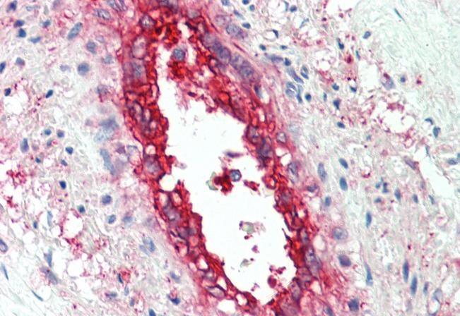
ARG63661 anti-EHD2 antibody IHC-P image
Immunohistochemistry: Paraffin-embedded Human vessel tissue. Antigen Retrieval: Steam tissue section in Citrate buffer (pH 6.0). The tissue section was stained with ARG63661 anti-EHD2 antibody at 5 µg/ml dilution followed by AP-staining.
-
ARG63661 anti-EHD2 antibody WB image
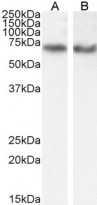
Western blot: 35 µg of A549 (A) and HeLa (B) cell lysates (in RIPA buffer) stained with ARG63661 anti-EHD2 antibody at 0.1 µg/ml (A) and 0.3 µg/ml (B) dilutions and incubated at RT for 1 hour.
-
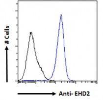
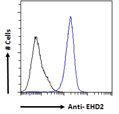
ARG63661 anti-EHD2 antibody FACS image
Flow Cytometry: Paraformaldehyde-fixed A431 cells permeabilized with 0.5% Triton. Cells were stained with ARG63661 anti-EHD2 antibody (blue line) at 10 µg/ml dilution for 1 hour, followed by incubation with Alexa FluorR 488 labelled secondary antibody. IgG control: Unimmunized goat IgG (black line).
-

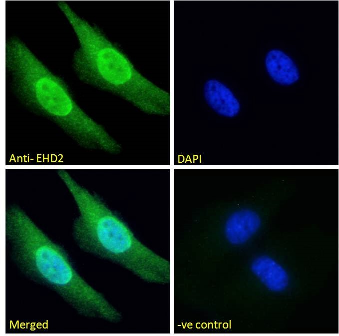
ARG63661 anti-EHD2 antibody ICC/IF image
Immunofluorescence: Paraformaldehyde fixed HeLa cells permeabilized with 0.15% Triton. Cells were stained with ARG63661 anti-EHD2 antibody (green) at 10 µg/ml dilution for 1 hour. DAPI (blue) for nuclear staining. Negative control: Unimmunized goat IgG (green) at 10 µg/ml dilution.
-
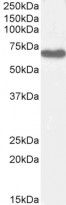
ARG63661 anti-EHD2 antibody WB image
Western blot: 35 µg of Human lung lysate (in RIPA buffer) stained with ARG63661 anti-EHD2 antibody at 0.3 µg/ml dilution and incubated at RT for 1 hour.