ARG42250
anti-EGFR antibody [EGFR1] (PE)
anti-EGFR antibody [EGFR1] (PE) for Flow cytometry and Human,Horse
Overview
| Product Description | PE-conjugated Mouse Monoclonal antibody [EGFR1] recognizes EGFR |
|---|---|
| Tested Reactivity | Hu, Hrs |
| Species Does Not React With | Ms |
| Tested Application | FACS |
| Specificity | The mouse monoclonal antibody EGFR1 reacts with extracellular domain of human protein kinase EGFR (ErbB1 / HER1); epitope within amino acids 6-273. |
| Host | Mouse |
| Clonality | Monoclonal |
| Clone | EGFR1 |
| Isotype | IgG2b, kappa |
| Target Name | EGFR |
| Antigen Species | Human |
| Immunogen | Human epidermoid carcinoma line A431. |
| Conjugation | PE |
| Alternate Names | PIG61; ERBB1; Proto-oncogene c-ErbB-1; Receptor tyrosine-protein kinase erbB-1; NISBD2; Epidermal growth factor receptor; ERBB; HER1; EC 2.7.10.1; mENA |
Application Instructions
| Application Suggestion |
|
||||
|---|---|---|---|---|---|
| Application Note | * The dilutions indicate recommended starting dilutions and the optimal dilutions or concentrations should be determined by the scientist. |
Properties
| Form | Liquid |
|---|---|
| Purification | Purified |
| Buffer | PBS and 15 mM Sodium azide. |
| Preservative | 15 mM Sodium azide |
| Storage Instruction | Aliquot and store in the dark at 2-8°C. Keep protected from prolonged exposure to light. Avoid repeated freeze/thaw cycles. Suggest spin the vial prior to opening. The antibody solution should be gently mixed before use. |
| Note | For laboratory research only, not for drug, diagnostic or other use. |
Bioinformation
| Database Links | |
|---|---|
| Gene Symbol | EGFR |
| Gene Full Name | epidermal growth factor receptor |
| Background | The protein encoded by this gene is a transmembrane glycoprotein that is a member of the protein kinase superfamily. This protein is a receptor for members of the epidermal growth factor family. EGFR is a cell surface protein that binds to epidermal growth factor. Binding of the protein to a ligand induces receptor dimerization and tyrosine autophosphorylation and leads to cell proliferation. Mutations in this gene are associated with lung cancer. [provided by RefSeq, Jun 2016] |
| Function | Receptor tyrosine kinase binding ligands of the EGF family and activating several signaling cascades to convert extracellular cues into appropriate cellular responses (PubMed:2790960, PubMed:10805725, PubMed:27153536). Known ligands include EGF, TGFA/TGF-alpha, AREG, epigen/EPGN, BTC/betacellulin, epiregulin/EREG and HBEGF/heparin-binding EGF (PubMed:2790960, PubMed:7679104, PubMed:8144591, PubMed:9419975, PubMed:15611079, PubMed:12297049, PubMed:27153536, PubMed:20837704). Ligand binding triggers receptor homo- and/or heterodimerization and autophosphorylation on key cytoplasmic residues. The phosphorylated receptor recruits adapter proteins like GRB2 which in turn activates complex downstream signaling cascades. Activates at least 4 major downstream signaling cascades including the RAS-RAF-MEK-ERK, PI3 kinase-AKT, PLCgamma-PKC and STATs modules (PubMed:27153536). May also activate the NF-kappa-B signaling cascade (PubMed:11116146). Also directly phosphorylates other proteins like RGS16, activating its GTPase activity and probably coupling the EGF receptor signaling to the G protein-coupled receptor signaling (PubMed:11602604). Also phosphorylates MUC1 and increases its interaction with SRC and CTNNB1/beta-catenin (PubMed:11483589). Plays a role in enhancing learning and memory performance (By similarity). Isoform 2 may act as an antagonist of EGF action. (Microbial infection) Acts as a receptor for hepatitis C virus (HCV) in hepatocytes and facilitates its cell entry. Mediates HCV entry by promoting the formation of the CD81-CLDN1 receptor complexes that are essential for HCV entry and by enhancing membrane fusion of cells expressing HCV envelope glycoproteins. [UniProt] |
| Cellular Localization | Cell membrane; Single-pass type I membrane protein. ER membrane; Single-pass type I membrane protein. Golgi apparatus membrane; Single-pass type I membrane protein. Nucleus membrane; Single-pass type I membrane protein. Endosome. Endosome membrane. Nucleus. Note=In response to EGF, translocated from the cell membrane to the nucleus via Golgi and ER. Endocytosed upon activation by ligand. Colocalized with GPER1 in the nucleus of estrogen agonist-induced CAF. Isoform 2: Secreted. [UniProt] |
| Calculated MW | 134 kDa |
| PTM | Phosphorylation at Ser-695 is partial and occurs only if Thr-693 is phosphorylated. Phosphorylation at Thr-678 and Thr-693 by PRKD1 inhibits EGF-induced MAPK8/JNK1 activation. Dephosphorylation by PTPRJ prevents endocytosis and stabilizes the receptor at the plasma membrane. Autophosphorylation at Tyr-1197 is stimulated by methylation at Arg-1199 and enhances interaction with PTPN6. Autophosphorylation at Tyr-1092 and/or Tyr-1110 recruits STAT3. Dephosphorylated by PTPN1 and PTPN2. Monoubiquitinated and polyubiquitinated upon EGF stimulation; which does not affect tyrosine kinase activity or signaling capacity but may play a role in lysosomal targeting. Polyubiquitin linkage is mainly through 'Lys-63', but linkage through 'Lys-48', 'Lys-11' and 'Lys-29' also occurs. Deubiquitination by OTUD7B prevents degradation. Ubiquitinated by RNF115 and RNF126 (By similarity). Methylated. Methylation at Arg-1199 by PRMT5 stimulates phosphorylation at Tyr-1197. [UniProt] |
Images (1) Click the Picture to Zoom In
-
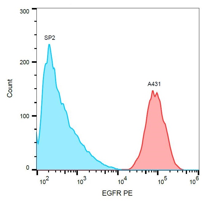
ARG42250 anti-EGFR antibody [EGFR1] (PE) FACS image
Flow Cytometry: A431 cells (red) and SP2 cells (negative sample, blue) stained with ARG42250 anti-EGFR antibody [EGFR1] (PE).