ARG54163
anti-EGFR antibody
anti-EGFR antibody for ICC/IF,Immunoprecipitation,Western blot and Human
Cancer antibody; Signaling Transduction antibody

Overview
| Product Description | Mouse Monoclonal antibody recognizes EGFR |
|---|---|
| Tested Reactivity | Hu |
| Tested Application | ICC/IF, IP, WB |
| Host | Mouse |
| Clonality | Monoclonal |
| Isotype | IgG1 |
| Target Name | EGFR |
| Antigen Species | Human |
| Immunogen | Purified recombinant human EGFR protein fragments expressed in E.coli. |
| Conjugation | Un-conjugated |
| Alternate Names | PIG61; ERBB1; Proto-oncogene c-ErbB-1; Receptor tyrosine-protein kinase erbB-1; NISBD2; Epidermal growth factor receptor; ERBB; HER1; EC 2.7.10.1; mENA |
Application Instructions
| Application Suggestion |
|
||||||||
|---|---|---|---|---|---|---|---|---|---|
| Application Note | IHC-P: Antigen Retrieval: Pressure cooking in Citrate buffer (pH 6.0). * The dilutions indicate recommended starting dilutions and the optimal dilutions or concentrations should be determined by the scientist. |
Properties
| Form | Liquid |
|---|---|
| Purification | Affinity purified |
| Buffer | PBS (pH 7.4), 0.02% Sodium azide and 50% Glycerol |
| Preservative | 0.02% Sodium azide |
| Stabilizer | 50% Glycerol |
| Concentration | 1.1 mg/ml |
| Storage Instruction | For continuous use, store undiluted antibody at 2-8°C for up to a week. For long-term storage, aliquot and store at -20°C. Storage in frost free freezers is not recommended. Avoid repeated freeze/thaw cycles. Suggest spin the vial prior to opening. The antibody solution should be gently mixed before use. |
| Note | For laboratory research only, not for drug, diagnostic or other use. |
Bioinformation
| Database Links | |
|---|---|
| Gene Symbol | EGFR |
| Background | EGFR is a transmembrane glycoprotein. It is a member of the protein kinase superfamily. This protein is a receptor for members of the epidermal growth factor family. EGFR is a cell surface protein that binds to epidermal growth factor. Binding of the protein to a ligand induces receptor dimerization and tyrosine autophosphorylation and leads to cell proliferation. Mutations in this gene are associated with lung cancer. [provided by RefSeq, Jun 2016] |
| Function | EGFR: Receptor tyrosine kinase binding ligands of the EGF family and activating several signaling cascades to convert extracellular cues into appropriate cellular responses (PubMed:2790960, PubMed:10805725, PubMed:27153536). Known ligands include EGF, TGFA/TGF-alpha, AREG, epigen/EPGN, BTC/betacellulin, epiregulin/EREG and HBEGF/heparin-binding EGF (PubMed:2790960, PubMed:7679104, PubMed:8144591, PubMed:9419975, PubMed:15611079, PubMed:12297049, PubMed:27153536, PubMed:20837704). Ligand binding triggers receptor homo- and/or heterodimerization and autophosphorylation on key cytoplasmic residues. The phosphorylated receptor recruits adapter proteins like GRB2 which in turn activates complex downstream signaling cascades. Activates at least 4 major downstream signaling cascades including the RAS-RAF-MEK-ERK, PI3 kinase-AKT, PLCgamma-PKC and STATs modules (PubMed:27153536). May also activate the NF-kappa-B signaling cascade (PubMed:11116146). Also directly phosphorylates other proteins like RGS16, activating its GTPase activity and probably coupling the EGF receptor signaling to the G protein-coupled receptor signaling (PubMed:11602604). Also phosphorylates MUC1 and increases its interaction with SRC and CTNNB1/beta-catenin (PubMed:11483589). Plays a role in enhancing learning and memory performance. Isoform 2 may act as an antagonist of EGF action. (Microbial infection) Acts as a receptor for hepatitis C virus (HCV) in hepatocytes and facilitates its cell entry. Mediates HCV entry by promoting the formation of the CD81-CLDN1 receptor complexes that are essential for HCV entry and by enhancing membrane fusion of cells expressing HCV envelope glycoproteins. [UniProt] |
| Cellular Localization | Cell membrane, Endoplasmic reticulum, Endosome, Golgi apparatus, Membrane, Nucleus, Secreted |
| Highlight | Related products: EGFR antibodies; EGFR Duos / Panels; Anti-Mouse IgG secondary antibodies; |
| Research Area | Cancer antibody; Signaling Transduction antibody |
| Calculated MW | 134 kDa |
| PTM | Phosphorylation at Ser-695 is partial and occurs only if Thr-693 is phosphorylated. Phosphorylation at Thr-678 and Thr-693 by PRKD1 inhibits EGF-induced MAPK8/JNK1 activation. Dephosphorylation by PTPRJ prevents endocytosis and stabilizes the receptor at the plasma membrane. Autophosphorylation at Tyr-1197 is stimulated by methylation at Arg-1199 and enhances interaction with PTPN6. Autophosphorylation at Tyr-1092 and/or Tyr-1110 recruits STAT3. Dephosphorylated by PTPN1 and PTPN2. Monoubiquitinated and polyubiquitinated upon EGF stimulation; which does not affect tyrosine kinase activity or signaling capacity but may play a role in lysosomal targeting. Polyubiquitin linkage is mainly through 'Lys-63', but linkage through 'Lys-48', 'Lys-11' and 'Lys-29' also occurs. Deubiquitination by OTUD7B prevents degradation. Ubiquitinated by RNF115 and RNF126 (By similarity). Methylated. Methylation at Arg-1199 by PRMT5 stimulates phosphorylation at Tyr-1197. |
Images (3) Click the Picture to Zoom In
-
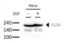
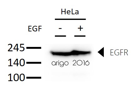
ARG54163 anti-EGFR antibody WB image
Western blot: 30 µg of HeLa untreated or treated with EGF and stained with ARG54163 anti-EGFR antibody at 1:1000 dilution.
-

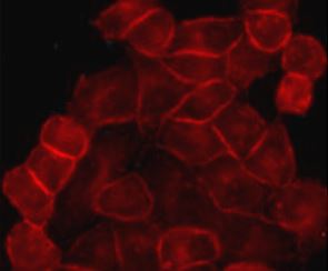
ARG54163 anti-EGFR antibody ICC/IF image
Immunofluorescence: HeLa cells stained with ARG54163 anti-EGFR antibody at 1:200 dilution.
-

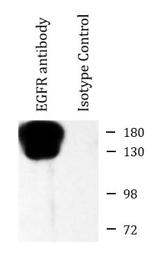
ARG54163 anti-EGFR antibody IP image
Immunoprecipitation: HeLa cell lysates were immunoprecipitated and stained with ARG54163 anti-EGFR antibody.
Customer's Feedback
 Good
Good

Review for anti-EGFR antibody
Application:IF/ICC
Sample:HeLa
Fixation Buffer:100% Methanol
Fixation Time:10 min
Fixation Temperature:RT ºC
Permeabilization Buffer:0.1% Triton X-100
Primary Antibody Dilution Factor:1:200
Primary Antibody Incubation Time:overnight
Primary Antibody Incubation Temperature:4 ºC
Conjugation of Secondary Antibody:FITC

 Excellent

Review for anti-EGFR antibody
Application:WB
Sample:HeLa untreated/treated with EGF
Sample Loading Amount:30 µg
Primary Antibody Dilution Factor:1:1000
Primary Antibody Incubation Time:overnight
Primary Antibody Incubation Temperature:4 ºC
Specific References
Poly (ethylene-co-vinyl alcohol) is a suitable substrate for human olfactory neuroepithelial cell differentiation in vitro through a defined regulatory pathway.
WB / Human


