ARG55685
anti-EBP1 antibody
anti-EBP1 antibody for Flow cytometry,IHC-Formalin-fixed paraffin-embedded sections,Western blot and Human,Mouse
Overview
| Product Description | Rabbit Polyclonal antibody recognizes EBP1 |
|---|---|
| Tested Reactivity | Hu, Ms |
| Tested Application | FACS, IHC-P, WB |
| Host | Rabbit |
| Clonality | Polyclonal |
| Isotype | IgG |
| Target Name | EBP1 |
| Antigen Species | Human |
| Immunogen | KLH-conjugated synthetic peptide corresponding to aa. 228-255 (Center) of Human EBP1. |
| Conjugation | Un-conjugated |
| Alternate Names | HG4-1; p38-2G4; Proliferation-associated protein 2G4; EBP1; ErbB3-binding protein 1; hG4-1; Cell cycle protein p38-2G4 homolog |
Application Instructions
| Application Suggestion |
|
||||||||
|---|---|---|---|---|---|---|---|---|---|
| Application Note | * The dilutions indicate recommended starting dilutions and the optimal dilutions or concentrations should be determined by the scientist. | ||||||||
| Positive Control | A2058 |
Properties
| Form | Liquid |
|---|---|
| Purification | This antibody is prepared by Saturated Ammonium Sulfate (SAS) precipitation followed by dialysis against PBS. |
| Buffer | PBS and 0.09% (W/V) Sodium azide. |
| Preservative | 0.09% (W/V) Sodium azide. |
| Storage Instruction | For continuous use, store undiluted antibody at 2-8°C for up to a week. For long-term storage, aliquot and store at -20°C or below. Storage in frost free freezers is not recommended. Avoid repeated freeze/thaw cycles. Suggest spin the vial prior to opening. The antibody solution should be gently mixed before use. |
| Note | For laboratory research only, not for drug, diagnostic or other use. |
Bioinformation
| Database Links |
Swiss-port # P50580 Mouse Proliferation-associated protein 2G4 Swiss-port # Q9UQ80 Human Proliferation-associated protein 2G4 |
|---|---|
| Gene Symbol | PA2G4 |
| Gene Full Name | proliferation-associated 2G4, 38kDa |
| Background | This gene encodes an RNA-binding protein that is involved in growth regulation. This protein is present in pre-ribosomal ribonucleoprotein complexes and may be involved in ribosome assembly and the regulation of intermediate and late steps of rRNA processing. This protein can interact with the cytoplasmic domain of the ErbB3 receptor and may contribute to transducing growth regulatory signals. This protein is also a transcriptional co-repressor of androgen receptor-regulated genes and other cell cycle regulatory genes through its interactions with histone deacetylases. This protein has been implicated in growth inhibition and the induction of differentiation of human cancer cells. Six pseudogenes, located on chromosomes 3, 6, 9, 18, 20 and X, have been identified. [provided by RefSeq, Jul 2008] |
| Function | May play a role in a ERBB3-regulated signal transduction pathway. Seems be involved in growth regulation. Acts a corepressor of the androgen receptor (AR) and is regulated by the ERBB3 ligand neuregulin-1/heregulin (HRG). Inhibits transcription of some E2F1-regulated promoters, probably by recruiting histone acetylase (HAT) activity. Binds RNA. Associates with 28S, 18S and 5.8S mature rRNAs, several rRNA precursors and probably U3 small nucleolar RNA. May be involved in regulation of intermediate and late steps of rRNA processing. May be involved in ribosome assembly. Mediates cap-independent translation of specific viral IRESs (internal ribosomal entry site) (By similarity). Regulates cell proliferation, differentiation, and survival. Isoform 1 suppresses apoptosis whereas isoform 2 promotes cell differentiation (By similarity). [UniProt] |
| Cellular Localization | Isoform 1: Cytoplasm Nucleus, nucleolus. Note=Tranlocates to the nucleus upon treatment with HRG. Phosphorylation at Ser-361 by PKC/PRKCD regulates its nucleolar localization |
| Calculated MW | 44 kDa |
| PTM | Phosphorylated on serine and threonine residues. Phosphorylation is enhanced by HRG treatment. Basal phosphorylation is PKC-dependent and HRG-induced phosphorylation is predominantly PKC-independent. Phosphorylation at Ser-361 by PKC/PRKCD regulates its nucleolar localization. In cancer cells, isoform 2 is polyubiquitinated leading to its proteasomal degradation and phosphorylation by PKC/PRKCD enhances polyubiquitination. |
Images (3) Click the Picture to Zoom In
-
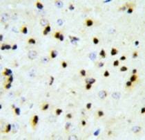
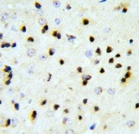
ARG55685 anti-EBP1 antibody IHC-P image
Immunohistochemistry: Formalin-fixed and paraffin-embedded Mouse brain tissue stained with ARG55685 anti-EBP1 antibody.
-
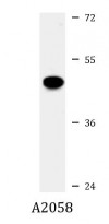
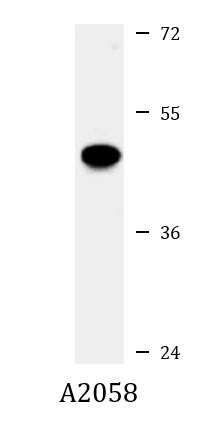
ARG55685 anti-EBP1 antibody WB image
Western blot: 35 µg of A2058 cell lysate stained with ARG55685 anti-EBP1 antibody.
-
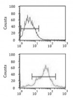
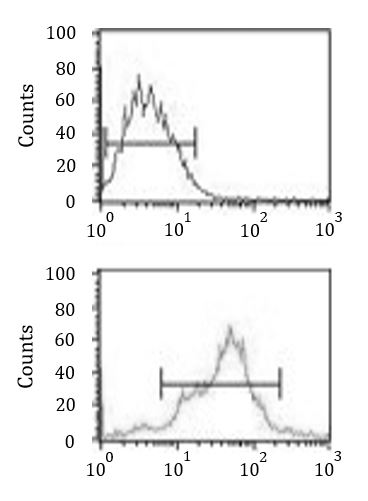
ARG55685 anti-EBP1 antibody FACS image
Flow Cytometry: MCF-7 cells stained with ARG55685 anti-EBP1 antibody (bottom histogram) or without primary antibody as control (top histogram), followed by incubation with FITC labelled secondary antibody.