ARG41292
anti-E Cadherin antibody [M168]
anti-E Cadherin antibody [M168] for ELISA,ICC/IF,IHC-Frozen sections,IHC-Formalin-fixed paraffin-embedded sections,Immunoprecipitation,Western blot and Human,Mouse,Rat,Horse
EMT Study antibody; Epithelial Marker antibody
Overview
| Product Description | Mouse Monoclonal antibody [M168] recognizes E Cadherin |
|---|---|
| Tested Reactivity | Hu, Ms, Rat, Hrs |
| Tested Application | ELISA, ICC/IF, IHC-Fr, IHC-P, IP, WB |
| Specificity | This antibody does not cross-react with VE-cadherin or N-cadherin. |
| Host | Mouse |
| Clonality | Monoclonal |
| Clone | M168 |
| Isotype | IgG1 |
| Target Name | E Cadherin |
| Antigen Species | Mouse |
| Immunogen | Recombinant protein around the C-terminus of Mouse E Cadherin. This sequence is highly conserved in Human and Rat E cadherin. |
| Conjugation | Un-conjugated |
| Alternate Names | Uvomorulin; Arc-1; Cadherin-1; E-cadherin; CDHE; CD antigen CD324; ECAD; CAM 120/80; LCAM; Epithelial cadherin; UVO; CD324 |
Application Instructions
| Application Suggestion |
|
||||||||||||||
|---|---|---|---|---|---|---|---|---|---|---|---|---|---|---|---|
| Application Note | WB: Antibody is suggested to be diluted in 5% skimmed milk/Tris buffer with 0.04% Tween20 and incubated for 1 hour at room temperature. * The dilutions indicate recommended starting dilutions and the optimal dilutions or concentrations should be determined by the scientist. |
||||||||||||||
| Observed Size | ~ 120 kDa |
Properties
| Form | Liquid |
|---|---|
| Purification | Purification with Protein A. |
| Buffer | PBS, 0.05% Sodium azide, 50% Glycerol and 1 mg/ml BSA. |
| Preservative | 0.05% Sodium azide |
| Stabilizer | 50% Glycerol and 1 mg/ml BSA |
| Storage Instruction | For continuous use, store undiluted antibody at 2-8°C for up to a week. For long-term storage, aliquot and store at -20°C. Storage in frost free freezers is not recommended. Avoid repeated freeze/thaw cycles. Suggest spin the vial prior to opening. The antibody solution should be gently mixed before use. |
| Note | For laboratory research only, not for drug, diagnostic or other use. |
Bioinformation
| Database Links | |
|---|---|
| Gene Symbol | CDH1 |
| Gene Full Name | cadherin 1, type 1 |
| Background | E Cadherin is a classical cadherin of the cadherin superfamily. Alternative splicing results in multiple transcript variants, at least one of which encodes a preproprotein that is proteolytically processed to generate the mature glycoprotein. This calcium-dependent cell-cell adhesion protein is comprised of five extracellular cadherin repeats, a transmembrane region and a highly conserved cytoplasmic tail. Mutations in this gene are correlated with gastric, breast, colorectal, thyroid and ovarian cancer. Loss of function of this gene is thought to contribute to cancer progression by increasing proliferation, invasion, and/or metastasis. The ectodomain of this protein mediates bacterial adhesion to mammalian cells and the cytoplasmic domain is required for internalization. This gene is present in a gene cluster with other members of the cadherin family on chromosome 16. [provided by RefSeq, Nov 2015] |
| Function | Cadherins are calcium-dependent cell adhesion proteins (PubMed:11976333). They preferentially interact with themselves in a homophilic manner in connecting cells; cadherins may thus contribute to the sorting of heterogeneous cell types. CDH1 is involved in mechanisms regulating cell-cell adhesions, mobility and proliferation of epithelial cells (PubMed:11976333). Has a potent invasive suppressor role. It is a ligand for integrin alpha-E/beta-7. E-Cad/CTF2 promotes non-amyloidogenic degradation of Abeta precursors. Has a strong inhibitory effect on APP C99 and C83 production. (Microbial infection) Serves as a receptor for Listeria monocytogenes; internalin A (InlA) binds to this protein and promotes uptake of the bacteria. [UniProt] |
| Cellular Localization | Cell junction. Cell membrane; Single-pass type I membrane protein. Endosome. Golgi apparatus, trans-Golgi network. [UniProt] |
| Research Area | EMT Study antibody; Epithelial Marker antibody |
| Calculated MW | 97 kDa |
| PTM | During apoptosis or with calcium influx, cleaved by a membrane-bound metalloproteinase (ADAM10), PS1/gamma-secretase and caspase-3 to produce fragments of about 38 kDa (E-CAD/CTF1), 33 kDa (E-CAD/CTF2) and 29 kDa (E-CAD/CTF3), respectively. Processing by the metalloproteinase, induced by calcium influx, causes disruption of cell-cell adhesion and the subsequent release of beta-catenin into the cytoplasm. The residual membrane-tethered cleavage product is rapidly degraded via an intracellular proteolytic pathway. Cleavage by caspase-3 releases the cytoplasmic tail resulting in disintegration of the actin microfilament system. The gamma-secretase-mediated cleavage promotes disassembly of adherens junctions. N-glycosylation at Asn-637 is essential for expression, folding and trafficking. Ubiquitinated by a SCF complex containing SKP2, which requires prior phosphorylation by CK1/CSNK1A1. Ubiquitinated by CBLL1/HAKAI, requires prior phosphorylation at Tyr-754. [UniProt] |
Images (1) Click the Picture to Zoom In
-

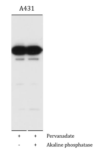
ARG41292 anti-E Cadherin antibody [M168] WB image
Western blot: A431 cells treated with pervanadate (1 mM) for 30 min (left) then treated with akaline phosphatase (right). The blots were stained with ARG41292 anti-E Cadherin antibody [M168].
Specific References
Mitochondrial citrate accumulation triggers senescence of alveolar epithelial cells contributing to pulmonary fibrosis in mice
IHC-P / Mouse


