ARG65402
anti-E Cadherin antibody [67A4]
anti-E Cadherin antibody [67A4] for Flow cytometry,ICC/IF,IHC-Frozen sections,Immunoprecipitation,Western blot and Human
EMT Study antibody; Epithelial Marker antibody

Overview
| Product Description | Mouse Monoclonal antibody [67A4] recognizes E Cadherin |
|---|---|
| Tested Reactivity | Hu |
| Tested Application | FACS, ICC/IF, IHC-Fr, IP, WB |
| Specificity | The mouse monoclonal antibody 67A4 recognizes CD324 / Ecadherin, an approximately 100 kDa epithelial cell adhesion molecule, whose detection is important for determination of invasive potential of epithelial neoplasms. HLDA VIII |
| Host | Mouse |
| Clonality | Monoclonal |
| Clone | 67A4 |
| Isotype | IgG1 |
| Target Name | E Cadherin |
| Immunogen | T-47D cells_x000D_ |
| Conjugation | Un-conjugated |
| Alternate Names | Uvomorulin; Arc-1; Cadherin-1; E-cadherin; CDHE; CD antigen CD324; ECAD; CAM 120/80; LCAM; Epithelial cadherin; UVO; CD324 |
Application Instructions
| Application Suggestion |
|
||||||||||||
|---|---|---|---|---|---|---|---|---|---|---|---|---|---|
| Application Note | * The dilutions indicate recommended starting dilutions and the optimal dilutions or concentrations should be determined by the scientist. | ||||||||||||
| Positive Control | FACS: CACO-2 and HT-29 IHC-Fr: Tonsil |
Properties
| Form | Liquid |
|---|---|
| Purification | Purified from cell culture supernatant by protein-A affinity chromatography. |
| Purity | > 95% (by SDS-PAGE) |
| Buffer | PBS (pH 7.4) and 15 mM Sodium azide |
| Preservative | 15 mM Sodium azide |
| Concentration | 1 mg/ml |
| Storage Instruction | For continuous use, store undiluted antibody at 2-8°C for up to a week. For long-term storage, aliquot and store at -20°C or below. Storage in frost free freezers is not recommended. Avoid repeated freeze/thaw cycles. Suggest spin the vial prior to opening. The antibody solution should be gently mixed before use. |
| Note | For laboratory research only, not for drug, diagnostic or other use. |
Bioinformation
| Database Links | |
|---|---|
| Gene Symbol | CDH1 |
| Gene Full Name | cadherin 1, type 1, E-cadherin (epithelial) |
| Background | E Cadherin is a classical cadherin of the cadherin superfamily. Alternative splicing results in multiple transcript variants, at least one of which encodes a preproprotein that is proteolytically processed to generate the mature glycoprotein. This calcium-dependent cell-cell adhesion protein is comprised of five extracellular cadherin repeats, a transmembrane region and a highly conserved cytoplasmic tail. Mutations in this gene are correlated with gastric, breast, colorectal, thyroid and ovarian cancer. Loss of function of this gene is thought to contribute to cancer progression by increasing proliferation, invasion, and/or metastasis. The ectodomain of this protein mediates bacterial adhesion to mammalian cells and the cytoplasmic domain is required for internalization. This gene is present in a gene cluster with other members of the cadherin family on chromosome 16. [provided by RefSeq, Nov 2015] |
| Function | Cadherins are calcium-dependent cell adhesion proteins (PubMed:11976333). They preferentially interact with themselves in a homophilic manner in connecting cells; cadherins may thus contribute to the sorting of heterogeneous cell types. CDH1 is involved in mechanisms regulating cell-cell adhesions, mobility and proliferation of epithelial cells (PubMed:11976333). Has a potent invasive suppressor role. It is a ligand for integrin alpha-E/beta-7. E-Cad/CTF2 promotes non-amyloidogenic degradation of Abeta precursors. Has a strong inhibitory effect on APP C99 and C83 production. (Microbial infection) Serves as a receptor for Listeria monocytogenes; internalin A (InlA) binds to this protein and promotes uptake of the bacteria. [UniProt] |
| Research Area | EMT Study antibody; Epithelial Marker antibody |
| Calculated MW | 97 kDa |
| PTM | During apoptosis or with calcium influx, cleaved by a membrane-bound metalloproteinase (ADAM10), PS1/gamma-secretase and caspase-3 to produce fragments of about 38 kDa (E-CAD/CTF1), 33 kDa (E-CAD/CTF2) and 29 kDa (E-CAD/CTF3), respectively. Processing by the metalloproteinase, induced by calcium influx, causes disruption of cell-cell adhesion and the subsequent release of beta-catenin into the cytoplasm. The residual membrane-tethered cleavage product is rapidly degraded via an intracellular proteolytic pathway. Cleavage by caspase-3 releases the cytoplasmic tail resulting in disintegration of the actin microfilament system. The gamma-secretase-mediated cleavage promotes disassembly of adherens junctions. N-glycosylation at Asn-637 is essential for expression, folding and trafficking. Ubiquitinated by a SCF complex containing SKP2, which requires prior phosphorylation by CK1/CSNK1A1. Ubiquitinated by CBLL1/HAKAI, requires prior phosphorylation at Tyr-754. |
Images (2) Click the Picture to Zoom In
-
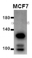
ARG65402 anti-E Cadherin antibody [67A4] WB image
Western blot: 20 µg of MCF7 cell lysate stained with ARG65402 anti-E Cadherin antibody [67A4] at 1:1000 dilution.
-
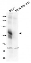
ARG65402 anti-E Cadherin antibody [67A4] WB image
Western blot: 20 µg of MCF7 and MDA-MB-231 cell lysates stained with ARG65402 anti-E Cadherin antibody [67A4] at 1:500 dilution.
MDA-MB-231 was a negative control.


Customer's Feedback
 Excellent

Review for anti-E Cadherin antibody [67A4]
Application:WB
Sample:MCF7
Sample Loading Amount:20 µg
Primary Antibody Dilution Factor:1:1000
Primary Antibody Incubation Time:overnight
Primary Antibody Incubation Temperature:4 ºC

 Average

Review for anti-E Cadherin antibody [67A4]
Application:WB
Sample:MCF7 and MDA-MB-231 (negative control)
Sample Loading Amount:20 µg
Primary Antibody Dilution Factor:1:500
Primary Antibody Incubation Time:overnight
Primary Antibody Incubation Temperature:4 ºC
Good

Review for anti-E Cadherin antibody [67A4]
Application:WB
Sample:MCF7
Sample Loading Amount:20 µg
Primary Antibody Dilution Factor:1:1000
Primary Antibody Incubation Time:overnight
Primary Antibody Incubation Temperature:4 ºC
Clone References
A complex 3D human tissue culture system based on mammary stromal cells and silk scaffolds for modeling breast morphogenesis and function.
ICC/IF / Human


Molecular events associated with epithelial to mesenchymal transition of nasopharyngeal carcinoma cells in the absence of Epstein-Barr virus genome.
Differential effects of allergens and irritants on early differentiating monocyte-derived dendritic cells.
Chronic renal allograft dysfunction: the role of T cell-mediated tubular epithelial to mesenchymal cell transition.


