ARG64961
anti-Decorin antibody
anti-Decorin antibody for IHC-Formalin-fixed paraffin-embedded sections,Western blot and Human
Developmental Biology antibody; Signaling Transduction antibody
Overview
| Product Description | Goat Polyclonal antibody recognizes Decorin |
|---|---|
| Tested Reactivity | Hu |
| Predict Reactivity | Cow, Pig |
| Tested Application | IHC-P, WB |
| Specificity | This antibody is expected to recognize the three reported isoforms a, b and c (NP_001911.1; NP_598011.1; NP_598012.1 resp.). Reported variants NP_598010.1 and NP_001911.1 represent identical protein. |
| Host | Goat |
| Clonality | Polyclonal |
| Isotype | IgG |
| Target Name | Decorin |
| Antigen Species | Human |
| Immunogen | C-KISRVDAASLKGLNN |
| Conjugation | Un-conjugated |
| Alternate Names | Bone proteoglycan II; PG40; PGII; CSCD; SLRR1B; PGS2; PG-S2; DSPG2; Decorin |
Application Instructions
| Application Suggestion |
|
||||||
|---|---|---|---|---|---|---|---|
| Application Note | WB: Recommend incubate at RT for 1h. IHC-P: Antigen Retrieval: Steam tissue section in Citrate buffer (pH 6.0). * The dilutions indicate recommended starting dilutions and the optimal dilutions or concentrations should be determined by the scientist. |
Properties
| Form | Liquid |
|---|---|
| Purification | Purified from goat serum by ammonium sulphate precipitation followed by antigen affinity chromatography using the immunizing peptide. |
| Buffer | Tris saline (pH 7.3), 0.02% Sodium azide and 0.5% BSA |
| Preservative | 0.02% Sodium azide |
| Stabilizer | 0.5% BSA |
| Concentration | 0.5 mg/ml |
| Storage Instruction | For continuous use, store undiluted antibody at 2-8°C for up to a week. For long-term storage, aliquot and store at -20°C or below. Storage in frost free freezers is not recommended. Avoid repeated freeze/thaw cycles. Suggest spin the vial prior to opening. The antibody solution should be gently mixed before use. |
| Note | For laboratory research only, not for drug, diagnostic or other use. |
Bioinformation
| Database Links | |
|---|---|
| Background | The protein encoded by this gene is a small cellular or pericellular matrix proteoglycan that is closely related in structure to biglycan protein. The encoded protein and biglycan are thought to be the result of a gene duplication. This protein is a component of connective tissue, binds to type I collagen fibrils, and plays a role in matrix assembly. It contains one attached glycosaminoglycan chain. This protein is capable of suppressing the growth of various tumor cell lines. There are multiple alternatively spliced transcript variants known for this gene. This gene is a candidate gene for Marfan syndrome. [provided by RefSeq, Jul 2008] |
| Research Area | Developmental Biology antibody; Signaling Transduction antibody |
| Calculated MW | 40 kDa |
| PTM | The attached glycosaminoglycan chain can be either chondroitin sulfate or dermatan sulfate depending upon the tissue of origin. |
Images (2) Click the Picture to Zoom In
-
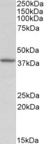
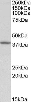
ARG64961 anti-Decorin antibody WB image
Western Blot: Human Kidney lysate (35 µg protein in RIPA buffer) stained with ARG64961 anti-Decorin antibody at 0.5 µg/ml dilution.
-
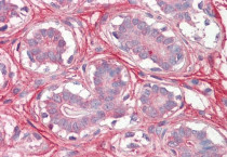
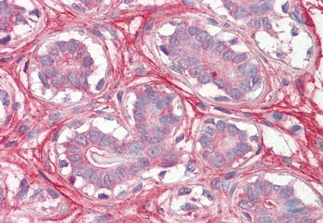
ARG64961 anti-Decorin antibody IHC-P image
Immunohistochemistry: Paraffin-embedded Human breast tissue. Antigen Retrieval: Steam tissue section in Citrate buffer (pH 6.0). The tissue section was stained with ARG64961 anti-Decorin antibody at 3.75 µg/ml dilution followed by AP-staining.