ARG62984
anti-Daxx antibody [DAXX-01]
anti-Daxx antibody [DAXX-01] for ICC/IF,Western blot and Human
Cell Biology and Cellular Response antibody; Cell Death antibody; Gene Regulation antibody; Microbiology and Infectious Disease antibody
Overview
| Product Description | Mouse Monoclonal antibody [DAXX-01] recognizes Daxx |
|---|---|
| Tested Reactivity | Hu |
| Tested Application | ICC/IF, WB |
| Specificity | The clone DAXX-01 reacts with Daxx, a death domain containing protein mainly expressed in fetal and adult human and mouse tissue. |
| Host | Mouse |
| Clonality | Monoclonal |
| Clone | DAXX-01 |
| Isotype | IgG1 |
| Target Name | Daxx |
| Antigen Species | Human |
| Immunogen | Recombinant C-terminal part (aa 558-740) of human Daxx. |
| Conjugation | Un-conjugated |
| Alternate Names | Daxx; ETS1-associated protein 1; hDaxx; Fas death domain-associated protein; Death domain-associated protein 6; DAP6; EAP1; BING2 |
Application Instructions
| Application Suggestion |
|
||||||
|---|---|---|---|---|---|---|---|
| Application Note | * The dilutions indicate recommended starting dilutions and the optimal dilutions or concentrations should be determined by the scientist. |
Properties
| Form | Liquid |
|---|---|
| Purification | Purified from ascites by protein-A affinity chromatography. |
| Purity | > 95% (by SDS-PAGE) |
| Buffer | PBS (pH 7.4) and 15 mM Sodium azide |
| Preservative | 15 mM Sodium azide |
| Concentration | 1 mg/ml |
| Storage Instruction | For continuous use, store undiluted antibody at 2-8°C for up to a week. For long-term storage, aliquot and store at -20°C or below. Storage in frost free freezers is not recommended. Avoid repeated freeze/thaw cycles. Suggest spin the vial prior to opening. The antibody solution should be gently mixed before use. |
| Note | For laboratory research only, not for drug, diagnostic or other use. |
Bioinformation
| Database Links | |
|---|---|
| Gene Symbol | DAXX |
| Gene Full Name | death-domain associated protein |
| Background | Daxx is an apoptosis-modulating death domain-associated protein with functions in transcriptional regulation. Daxx functions bot in cytoplasm, where it interacts with Fas, and in nucleus (residing in PML oncogenic domains), where it is involved in SUMO-dependent regulation of transcription and subnuclear compartmentalization. Daxx senzitizes the cells to apoptosis, but on the other hand, this protein may also serve in preventing apoptosis in the early embryo. Even regarding the transcription, Daxx can serve both as a corepressor and a coactivator. During ischemic stress, Daxx translocates from the nucleus to the cytoplasm, where in regulates sodium hydrogen exchanger isoform 1 (NHE1)._x000D_ |
| Function | Transcription corepressor known to repress transcriptional potential of several sumoylated transcription factors. Down-regulates basal and activated transcription. Its transcription repressor activity is modulated by recruiting it to subnuclear compartments like the nucleolus or PML/POD/ND10 nuclear bodies through interactions with MCSR1 and PML, respectively. Seems to regulate transcription in PML/POD/ND10 nuclear bodies together with PML and may influence TNFRSF6-dependent apoptosis thereby. Inhibits transcriptional activatiopn of PAX3 and ETS1 through direct protein-protein interactions. Modulates PAX5 activity; the function seems to involve CREBBP. Acts as an adapter protein in a MDM2-DAXX-USP7 complex by regulating the RING-finger E3 ligase MDM2 ubiquitination activity. Under non-stress condition, in association with the deubiquitinating USP7, prevents MDM2 self-ubiquitination and enhances the intrinsic E3 ligase activity of MDM2 towards TP53, thereby promoting TP53 ubiquitination and subsequent proteasomal degradation. Upon DNA damage, its association with MDM2 and USP7 is disrupted, resulting in increased MDM2 autoubiquitination and consequently, MDM2 degradation, which leads to TP53 stabilization. Acts as histone chaperone that facilitates deposition of histone H3.3. Acts as targeting component of the chromatin remodeling complex ATRX:DAXX which has ATP-dependent DNA translocase activity and catalyzes the replication-independent deposition of histone H3.3 in pericentric DNA repeats outside S-phase and telomeres, and the in vitro remodeling of H3.3-containing nucleosomes. Does not affect the ATPase activity of ATRX but alleviates its transcription repression activity. Upopn neuronal activation asociates with regulatory elements of selected immediate early genes where it promotes deposition of histone H3.3 which may be linked to transcriptional induction of these genes. Required for the recruitment of histone H3.3:H4 dimers to PML-nuclear bodies (PML-NBs); the process is independent of ATRX and facilitated by ASF1A; PML-NBs are suggested to function as regulatory sites for the incorporation of newly synthesized histone H3.3 into chromatin. In case of overexpression of centromeric histone variant CENPA (as found in various tumors) is involved in its mislocalization to chromosomes; the ectopic localization involves a heterotypic tetramer containing CENPA, and histones H3.3 and H4 and decreases binding of CTCF to chromatin. Proposed to mediate activation of the JNK pathway and apoptosis via MAP3K5 in response to signaling from TNFRSF6 and TGFBR2. Interaction with HSPB1/HSP27 may prevent interaction with TNFRSF6 and MAP3K5 and block DAXX-mediated apoptosis. In contrast, in lymphoid cells JNC activation and TNFRSF6-mediated apoptosis may not involve DAXX. Shows restriction activity towards human cytomegalovirus (HCMV). [UniProt] |
| Research Area | Cell Biology and Cellular Response antibody; Cell Death antibody; Gene Regulation antibody; Microbiology and Infectious Disease antibody |
| Calculated MW | 81 kDa |
| PTM | Sumoylated with SUMO1 on multiple lysine residues. Phosphorylated by HIPK1 upon glucose deprivation. Polyubiquitinated; which is promoted by CUL3 and SPOP and results in proteasomal degradation. Ubiquitinated by MDM2; inducing its degradation. Deubiquitinated by USP7; leading to stabilize it. |
Images (2) Click the Picture to Zoom In
-

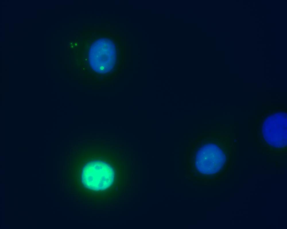
ARG62984 anti-Daxx antibody [DAXX-01] ICC/IF image
Immunofluorescence: HeLa cells transfected with pCDNA3-MycDaxx stained with ARG62984 anti-Daxx antibody [DAXX-01] (green)
Nuclei were stained with DAPI (blue). -

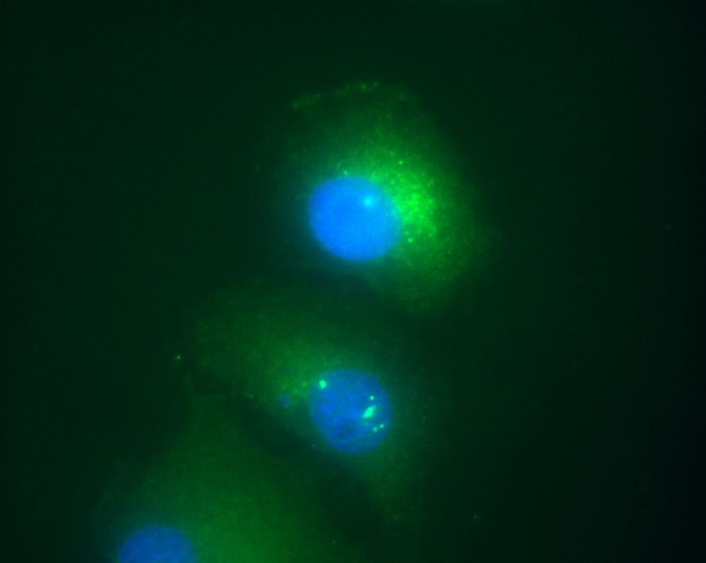
ARG62984 anti-Daxx antibody [DAXX-01] ICC/IF image
Immunofluorescence: HeLa cells co-transfected with pCDNA3-MycDaxx and pCDNA3-ASK1HA stained with ARG62984 anti-Daxx antibody [DAXX-01] (green)
Nuclei were stained with DAPI (blue). Note the translocation of Daxx from nucleus to cytoplasm.
Clone References
PML nuclear bodies are highly organised DNA-protein structures with a function in heterochromatin remodelling at the G2 phase.


