ARG54848
anti-DYRK2 antibody
anti-DYRK2 antibody for Western blot and Human
Signaling Transduction antibody
Overview
| Product Description | Mouse Monoclonal antibody recognizes DYRK2 |
|---|---|
| Tested Reactivity | Hu |
| Tested Application | WB |
| Host | Mouse |
| Clonality | Monoclonal |
| Clone | 492CT4.2.4 |
| Isotype | IgG1 |
| Target Name | DYRK2 |
| Antigen Species | Human |
| Immunogen | KLH-conjugated synthetic peptide corresponding to aa. 105-135 of Human DYRK2. |
| Conjugation | Un-conjugated |
| Alternate Names | Dual specificity tyrosine-phosphorylation-regulated kinase 2; EC 2.7.12.1 |
Application Instructions
| Application Suggestion |
|
||||
|---|---|---|---|---|---|
| Application Note | * The dilutions indicate recommended starting dilutions and the optimal dilutions or concentrations should be determined by the scientist. | ||||
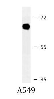
| Positive Control | A549 |
Properties
| Form | Liquid |
|---|---|
| Purification | This antibody is prepared by Saturated Ammonium Sulfate (SAS) precipitation followed by dialysis against PBS. |
| Buffer | PBS and 0.09% (W/V) Sodium azide |
| Preservative | 0.09% (W/V) Sodium azide |
| Storage Instruction | For continuous use, store undiluted antibody at 2-8°C for up to a week. For long-term storage, aliquot and store at -20°C or below. Storage in frost free freezers is not recommended. Avoid repeated freeze/thaw cycles. Suggest spin the vial prior to opening. The antibody solution should be gently mixed before use. |
| Note | For laboratory research only, not for drug, diagnostic or other use. |
Bioinformation
| Database Links |
Swiss-port # Q92630 Human Dual specificity tyrosine-phosphorylation-regulated kinase 2 |
|---|---|
| Gene Symbol | DYRK2 |
| Gene Full Name | dual-specificity tyrosine-(Y)-phosphorylation regulated kinase 2 |
| Background | DYRK2 belongs to a family of protein kinases whose members are presumed to be involved in cellular growth and/or development. The family is defined by structural similarity of their kinase domains and their capability to autophosphorylate on tyrosine residues. DYRK2 has demonstrated tyrosine autophosphorylation and catalyzed phosphorylation of histones H3 and H2B in vitro. Two isoforms of DYRK2 have been isolated. The predominant isoform, isoform 1, lacks a 5' terminal insert. [provided by RefSeq, Jul 2008] |
| Function | Serine/threonine-protein kinase involved in the regulation of the mitotic cell cycle, cell proliferation, apoptosis, organization of the cytoskeleton and neurite outgrowth. Functions in part via its role in ubiquitin-dependent proteasomal protein degradation. Functions downstream of ATM and phosphorylates p53/TP53 at 'Ser-46', and thereby contributes to the induction of apoptosis in response to DNA damage. Phosphorylates NFATC1, and thereby inhibits its accumulation in the nucleus and its transcription factor activity. Phosphorylates EIF2B5 at 'Ser-544', enabling its subsequent phosphorylation and inhibition by GSK3B. Likewise, phosphorylation of NFATC1, CRMP2/DPYSL2 and CRMP4/DPYSL3 promotes their subsequent phosphorylation by GSK3B. May play a general role in the priming of GSK3 substrates. Inactivates GYS1 by phosphorylation at 'Ser-641', and potentially also a second phosphorylation site, thus regulating glycogen synthesis. Mediates EDVP E3 ligase complex formation and is required for the phosphorylation and subsequent degradation of KATNA1. Phosphorylates TERT at 'Ser-457', promoting TERT ubiquitination by the EDVP complex. Phosphorylates SIAH2, and thereby increases its ubiquitin ligase activity. Promotes the proteasomal degradation of MYC and JUN, and thereby regulates progress through the mitotic cell cycle and cell proliferation. Promotes proteasomal degradation of GLI2 and GLI3, and thereby plays a role in smoothened and sonic hedgehog signaling. Plays a role in cytoskeleton organization and neurite outgrowth via its phosphorylation of DCX and DPYSL2. Phosphorylates CRMP2/DPYSL2, CRMP4/DPYSL3, DCX, EIF2B5, EIF4EBP1, GLI2, GLI3, GYS1, JUN, MDM2, MYC, NFATC1, p53/TP53, TAU/MAPT and KATNA1. Can phosphorylate histone H1, histone H3 and histone H2B (in vitro). Can phosphorylate CARHSP1 (in vitro). [UniProt] |
| Cellular Localization | Cytoplasm. Nucleus. Note=Translocates into the nucleus following DNA damage |
| Research Area | Signaling Transduction antibody |
| Calculated MW | 67 kDa |
| PTM | Autophosphorylates cotranslationally on the second tyrosine residue in the Tyr-X-Tyr motif in the activation loop, but once mature, does not have any protein tyrosine kinase activity. Phosphorylated at Thr-106 and Ser-442 by ATM in response to genotoxic stress. Under normal conditions, polyubiquitinated in the nucleus by MDM2, leading to its proteasomal degradation. Phosphorylation on Thr-106 and Ser-442 by ATM in response to genotoxic stress disrupts MDM2 binding and prevents MDM2-mediated ubiquitination and subsequent proteasomal degradation. Polyubiquitinated by SIAH2, leading to its proteasomal degradation. Polyubiquitinated by SIAH2 occurs under normal conditions, and is enhanced in response to hypoxia. |
Images (1) Click the Picture to Zoom In
-
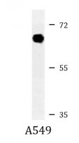
ARG54848 anti-DYRK2 antibody WB image
Western blot: 35 µg of A549 cell lysate stained with ARG54848 anti-DYRK2 antibody.