ARG63167
anti-DUSP1 antibody
anti-DUSP1 antibody for IHC-Formalin-fixed paraffin-embedded sections,Western blot and Human
Signaling Transduction antibody
Overview
| Product Description | Goat Polyclonal antibody recognizes DUSP1 |
|---|---|
| Tested Reactivity | Hu |
| Predict Reactivity | Ms, Rat, Cow, Dog |
| Tested Application | IHC-P, WB |
| Host | Goat |
| Clonality | Polyclonal |
| Isotype | IgG |
| Target Name | DUSP1 |
| Antigen Species | Human |
| Immunogen | SYLQSPITTSPSC |
| Conjugation | Un-conjugated |
| Alternate Names | PTPN10; MKP-1; MKP1; MAP kinase phosphatase 1; CL100; EC 3.1.3.16; Mitogen-activated protein kinase phosphatase 1; HVH1; Dual specificity protein phosphatase hVH1; Dual specificity protein phosphatase 1; EC 3.1.3.48; Protein-tyrosine phosphatase CL100 |
Application Instructions
| Application Suggestion |
|
||||||
|---|---|---|---|---|---|---|---|
| Application Note | WB: Recommend incubate at RT for 1h. IHC-P: Antigen Retrieval: Heat mediation was performed in Citrate buffer (pH 6.0). * The dilutions indicate recommended starting dilutions and the optimal dilutions or concentrations should be determined by the scientist. |
Properties
| Form | Liquid |
|---|---|
| Purification | Purified from goat serum by antigen affinity chromatography. |
| Buffer | Tris saline (pH 7.3), 0.02% Sodium azide and 0.5% BSA. |
| Preservative | 0.02% Sodium azide |
| Stabilizer | 0.5% BSA |
| Concentration | 0.5 mg/ml |
| Storage Instruction | For continuous use, store undiluted antibody at 2-8°C for up to a week. For long-term storage, aliquot and store at -20°C or below. Storage in frost free freezers is not recommended. Avoid repeated freeze/thaw cycles. Suggest spin the vial prior to opening. The antibody solution should be gently mixed before use. |
| Note | For laboratory research only, not for drug, diagnostic or other use. |
Bioinformation
| Database Links |
Swiss-port # P28562 Human Dual specificity protein phosphatase 1 |
|---|---|
| Background | The expression of DUSP1 gene is induced in human skin fibroblasts by oxidative/heat stress and growth factors. It specifies a protein with structural features similar to members of the non-receptor-type protein-tyrosine phosphatase family, and which has significant amino-acid sequence similarity to a Tyr/Ser-protein phosphatase encoded by the late gene H1 of vaccinia virus. The bacterially expressed and purified DUSP1 protein has intrinsic phosphatase activity, and specifically inactivates mitogen-activated protein (MAP) kinase in vitro by the concomitant dephosphorylation of both its phosphothreonine and phosphotyrosine residues. Furthermore, it suppresses the activation of MAP kinase by oncogenic ras in extracts of Xenopus oocytes. Thus, DUSP1 may play an important role in the human cellular response to environmental stress as well as in the negative regulation of cellular proliferation. [provided by RefSeq, Jul 2008] |
| Research Area | Signaling Transduction antibody |
| Calculated MW | 39 kDa |
| PTM | Phosphorylation at Ser-359 and Ser-364 by MAPK1/ERK2 and MAPK3/ERK1 reduces its rate of degradation. |
Images (2) Click the Picture to Zoom In
-

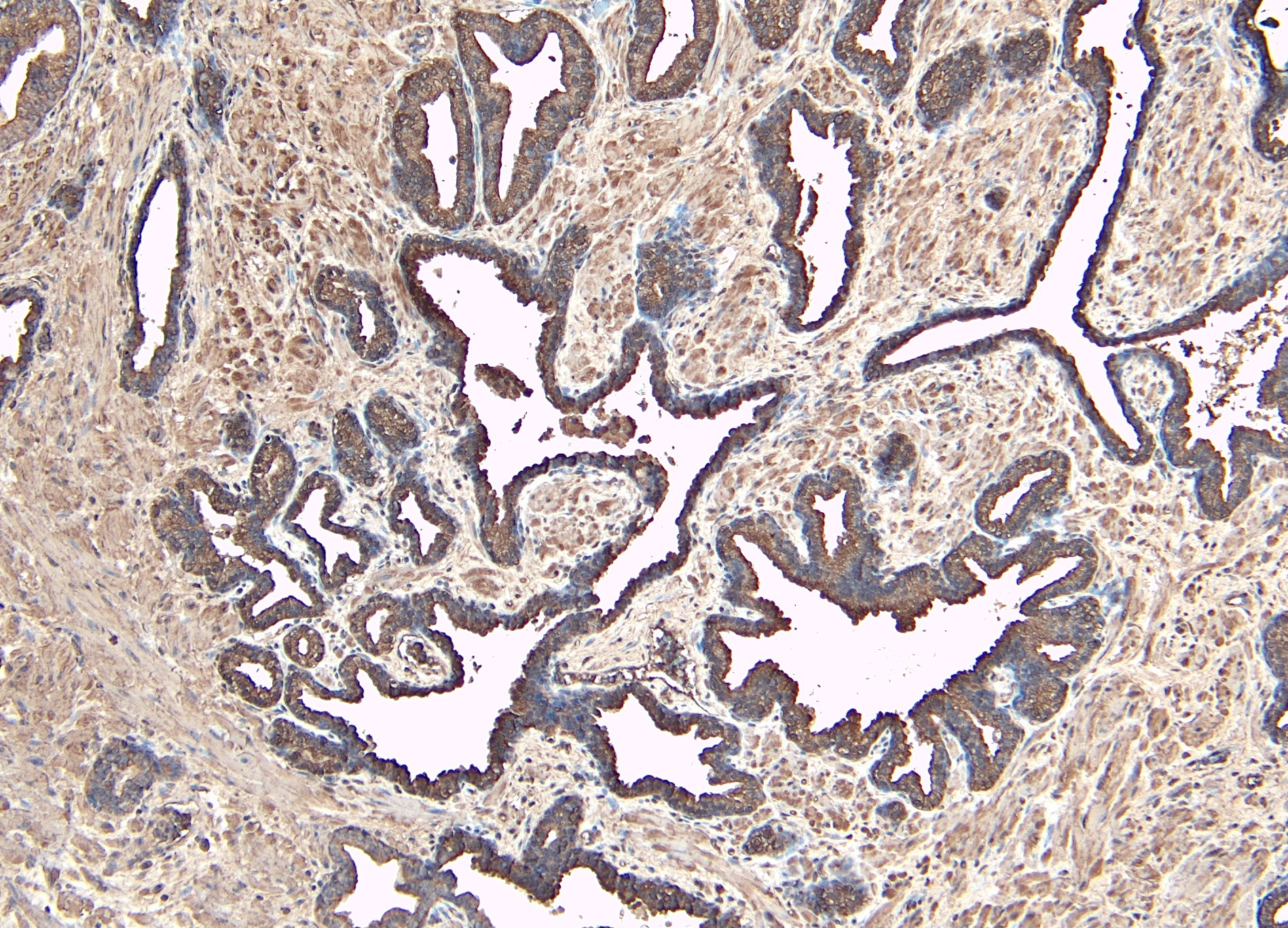
ARG63167 anti-DUSP1 antibody IHC-P image
Immunohistochemistry: Paraffin-embedded Human prostate tissue. Antigen Retrieval: Heat mediation was performed in Citrate buffer (pH 6.0). The tissue section was stained with ARG63167 anti-DUSP1 antibody at 8 µg/ml dilution followed by HRP-staining.
-

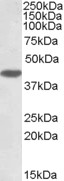
ARG63167 anti-DUSP1 antibody WB image
Western blot: 35 µg of HeLa cell lysate (in RIPA buffer) stained with ARG63167 anti-DUSP1 antibody at 1 µg/ml dilution and incubated at RT for 1 hour.