ARG40509
anti-DDR2 antibody
anti-DDR2 antibody for IHC-Formalin-fixed paraffin-embedded sections and Mouse,Rat
Overview
| Product Description | Rabbit Polyclonal antibody recognizes DDR2 |
|---|---|
| Tested Reactivity | Ms, Rat |
| Tested Application | IHC-P |
| Host | Rabbit |
| Clonality | Polyclonal |
| Isotype | IgG |
| Target Name | DDR2 |
| Antigen Species | Human |
| Immunogen | Recombinant protein corresponding to T801-E855 of Human DDR2. |
| Conjugation | Un-conjugated |
| Alternate Names | Tyrosine-protein kinase TYRO10; Receptor protein-tyrosine kinase TKT; Neurotrophic tyrosine kinase, receptor-related 3; MIG20a; Discoidin domain-containing receptor tyrosine kinase 2; Discoidin domain receptor 2; TKT; CD antigen CD167b; NTRKR3; CD167 antigen-like family member B; EC 2.7.10.1; TYRO10; Discoidin domain-containing receptor 2 |
Application Instructions
| Application Suggestion |
|
||||
|---|---|---|---|---|---|
| Application Note | IHC-P: Antigen Retrieval: Heat mediation was performed in Citrate buffer (pH 6.0) for 20 min. * The dilutions indicate recommended starting dilutions and the optimal dilutions or concentrations should be determined by the scientist. |
Properties
| Form | Liquid |
|---|---|
| Purification | Affinity purification with immunogen. |
| Buffer | 0.2% Na2HPO4, 0.9% NaCl, 0.05% Sodium azide and 4% Trehalose. |
| Preservative | 0.05% Sodium azide |
| Stabilizer | 4% Trehalose |
| Concentration | 0.5 mg/ml |
| Storage Instruction | For continuous use, store undiluted antibody at 2-8°C for up to a week. For long-term storage, aliquot and store at -20°C or below. Storage in frost free freezers is not recommended. Avoid repeated freeze/thaw cycles. Suggest spin the vial prior to opening. The antibody solution should be gently mixed before use. |
| Note | For laboratory research only, not for drug, diagnostic or other use. |
Bioinformation
| Database Links |
Swiss-port # Q62371 Mouse Discoidin domain-containing receptor 2 |
|---|---|
| Gene Symbol | DDR2 |
| Gene Full Name | discoidin domain receptor tyrosine kinase 2 |
| Background | Receptor tyrosine kinases (RTKs) play a key role in the communication of cells with their microenvironment. These molecules are involved in the regulation of cell growth, differentiation, and metabolism. In several cases the biochemical mechanism by which RTKs transduce signals across the membrane has been shown to be ligand induced receptor oligomerization and subsequent intracellular phosphorylation. This autophosphorylation leads to phosphorylation of cytosolic targets as well as association with other molecules, which are involved in pleiotropic effects of signal transduction. RTKs have a tripartite structure with extracellular, transmembrane, and cytoplasmic regions. This gene encodes a member of a novel subclass of RTKs and contains a distinct extracellular region encompassing a factor VIII-like domain. Alternative splicing in the 5' UTR results in multiple transcript variants encoding the same protein. [provided by RefSeq, Jul 2008] |
| Function | Tyrosine kinase that functions as cell surface receptor for fibrillar collagen and regulates cell differentiation, remodeling of the extracellular matrix, cell migration and cell proliferation. Required for normal bone development. Regulates osteoblast differentiation and chondrocyte maturation via a signaling pathway that involves MAP kinases and leads to the activation of the transcription factor RUNX2. Regulates remodeling of the extracellular matrix by up-regulation of the collagenases MMP1, MMP2 and MMP13, and thereby facilitates cell migration and tumor cell invasion. Promotes fibroblast migration and proliferation, and thereby contributes to cutaneous wound healing. [UniProt] |
| Cellular Localization | Cell membrane; Single-pass type I membrane protein. [UniProt] |
| Calculated MW | 97 kDa |
| PTM | N-glycosylated. Tyrosine phosphorylated in response to collagen binding. Phosphorylated by SRC; this is required for activation and subsequent autophosphorylation on additional tyrosine residues. [UniProt] |
Images (2) Click the Picture to Zoom In
-

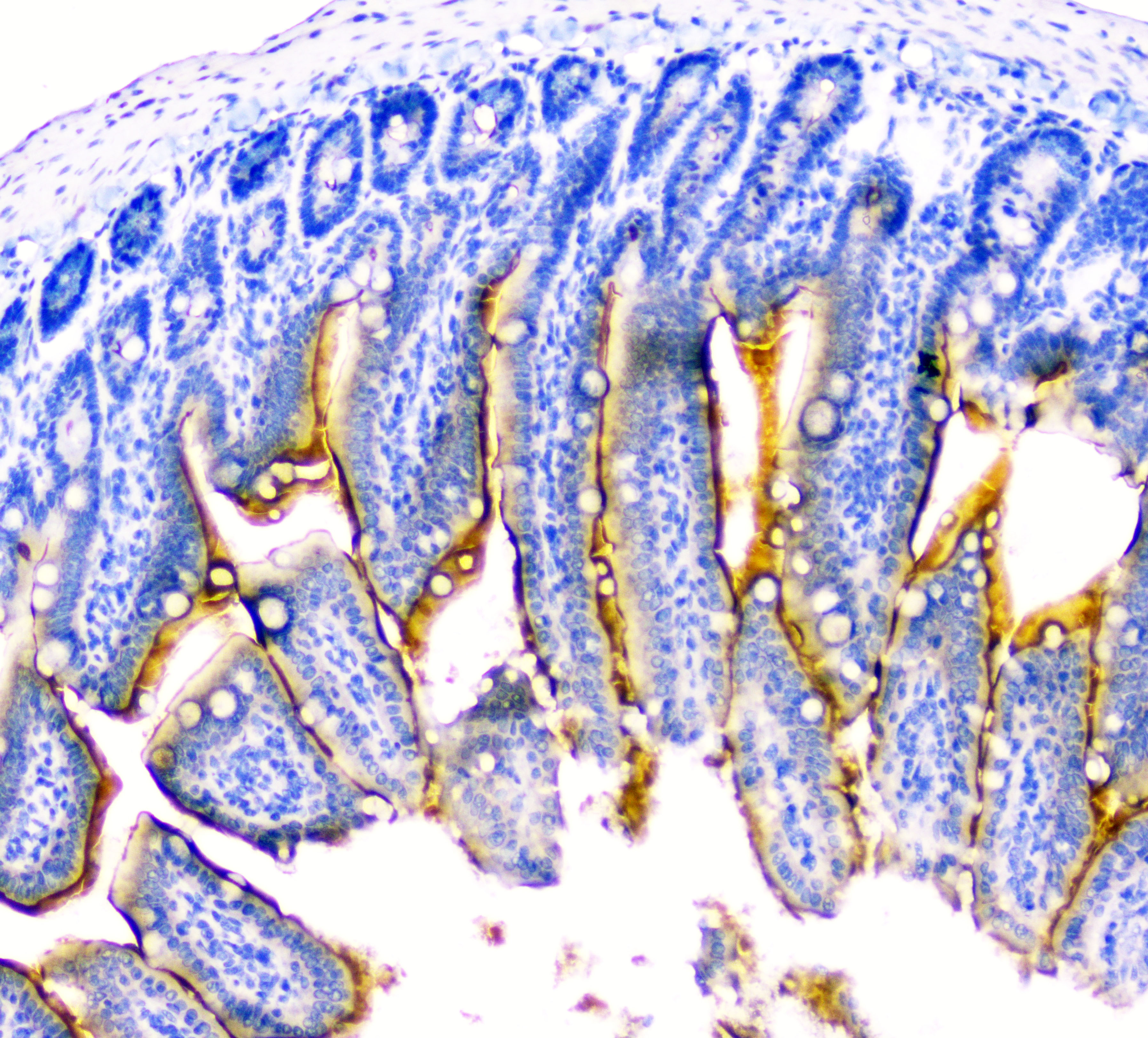
ARG40509 anti-DDR2 antibody IHC-P image
Immunohistochemistry: Paraffin-embedded Mouse small intestine tissue. Antigen Retrieval: Heat mediation was performed in Citrate buffer (pH 6.0, epitope retrieval solution) for 20 min. The tissue section was blocked with 10% goat serum. The tissue section was then stained with ARG40509 anti-DDR2 antibody at 2 µg/ml, overnight at 4°C.
-
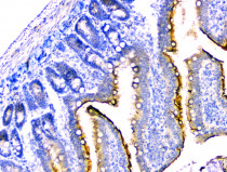
ARG40509 anti-DDR2 antibody IHC-P image
Immunohistochemistry: Paraffin-embedded Rat small intestine tissue. Antigen Retrieval: Heat mediation was performed in Citrate buffer (pH 6.0, epitope retrieval solution) for 20 min. The tissue section was blocked with 10% goat serum. The tissue section was then stained with ARG40509 anti-DDR2 antibody at 2 µg/ml, overnight at 4°C.