ARG58523
anti-DDB2 antibody
anti-DDB2 antibody for Flow cytometry,ICC/IF,Western blot and Human
Overview
| Product Description | Rabbit Polyclonal antibody recognizes DDB2 |
|---|---|
| Tested Reactivity | Hu |
| Tested Application | FACS, ICC/IF, WB |
| Host | Rabbit |
| Clonality | Polyclonal |
| Isotype | IgG |
| Target Name | DDB2 |
| Antigen Species | Human |
| Immunogen | Human DDB2 recombinant protein (Position: M1-T115). Human DDB2 shares 58.3% amino acid (aa) sequence identity with Mouse DDB2. |
| Conjugation | Un-conjugated |
| Alternate Names | DDBb; Damage-specific DNA-binding protein 2; DDBB; UV-damaged DNA-binding protein 2; DDB p48 subunit; UV-DDB2; UV-DDB 2; DNA damage-binding protein 2 |
Application Instructions
| Application Suggestion |
|
||||||||
|---|---|---|---|---|---|---|---|---|---|
| Application Note | * The dilutions indicate recommended starting dilutions and the optimal dilutions or concentrations should be determined by the scientist. |
Properties
| Form | Liquid |
|---|---|
| Purification | Affinity purification with immunogen. |
| Buffer | 0.9% NaCl, 0.2% Na2HPO4, 0.05% Sodium azide and 5% BSA. |
| Preservative | 0.05% Sodium azide |
| Stabilizer | 5% BSA |
| Concentration | 0.5 mg/ml |
| Storage Instruction | For continuous use, store undiluted antibody at 2-8°C for up to a week. For long-term storage, aliquot and store at -20°C or below. Storage in frost free freezers is not recommended. Avoid repeated freeze/thaw cycles. Suggest spin the vial prior to opening. The antibody solution should be gently mixed before use. |
| Note | For laboratory research only, not for drug, diagnostic or other use. |
Bioinformation
| Database Links | |
|---|---|
| Gene Symbol | DDB2 |
| Gene Full Name | damage-specific DNA binding protein 2, 48kDa |
| Background | This gene encodes a protein that is necessary for the repair of ultraviolet light-damaged DNA. This protein is the smaller subunit of a heterodimeric protein complex that participates in nucleotide excision repair, and this complex mediates the ubiquitylation of histones H3 and H4, which facilitates the cellular response to DNA damage. This subunit appears to be required for DNA binding. Mutations in this gene cause xeroderma pigmentosum complementation group E, a recessive disease that is characterized by an increased sensitivity to UV light and a high predisposition for skin cancer development, in some cases accompanied by neurological abnormalities. Two transcript variants encoding different isoforms have been found for this gene. [provided by RefSeq, Jul 2014] |
| Function | Required for DNA repair. Binds to DDB1 to form the UV-damaged DNA-binding protein complex (the UV-DDB complex). The UV-DDB complex may recognize UV-induced DNA damage and recruit proteins of the nucleotide excision repair pathway (the NER pathway) to initiate DNA repair. The UV-DDB complex preferentially binds to cyclobutane pyrimidine dimers (CPD), 6-4 photoproducts (6-4 PP), apurinic sites and short mismatches. Also appears to function as the substrate recognition module for the DCX (DDB1-CUL4-X-box) E3 ubiquitin-protein ligase complex DDB1-CUL4-ROC1 (also known as CUL4-DDB-ROC1 and CUL4-DDB-RBX1). The DDB1-CUL4-ROC1 complex may ubiquitinate histone H2A, histone H3 and histone H4 at sites of UV-induced DNA damage. The ubiquitination of histones may facilitate their removal from the nucleosome and promote subsequent DNA repair. The DDB1-CUL4-ROC1 complex also ubiquitinates XPC, which may enhance DNA-binding by XPC and promote NER. Isoform D1 and isoform D2 inhibit UV-damaged DNA repair. [UniProt] |
| Cellular Localization | Nucleus. Accumulates at sites of DNA damage following UV irradiation. [UniProt] |
| Calculated MW | 48 kDa |
| PTM | Phosphorylation by ABL1 negatively regulate UV-DDB activity. Ubiquitinated by CUL4A in response to UV irradiation. Ubiquitination appears to both impair DNA-binding and promotes ubiquitin-dependent proteolysis. Degradation of DDB2 at sites of DNA damage may be a prerequisite for their recognition by XPC and subsequent repair. CUL4A-mediated degradation appears to be promoted by ABL1. Ubiquitinated, leading to proteasomal degradation, and deubiquitinated by USP24. [UniProt] |
Images (3) Click the Picture to Zoom In
-
ARG58523 anti-DDB2 antibody ICC/IF image
Immunofluorescence: U2OS cells were blocked with 10% goat serum and then stained with ARG58523 anti-DDB2 antibody (red) at 2 µg/ml dilution, overnight at 4°C. DAPI (blue) for nuclear staining.
-
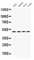
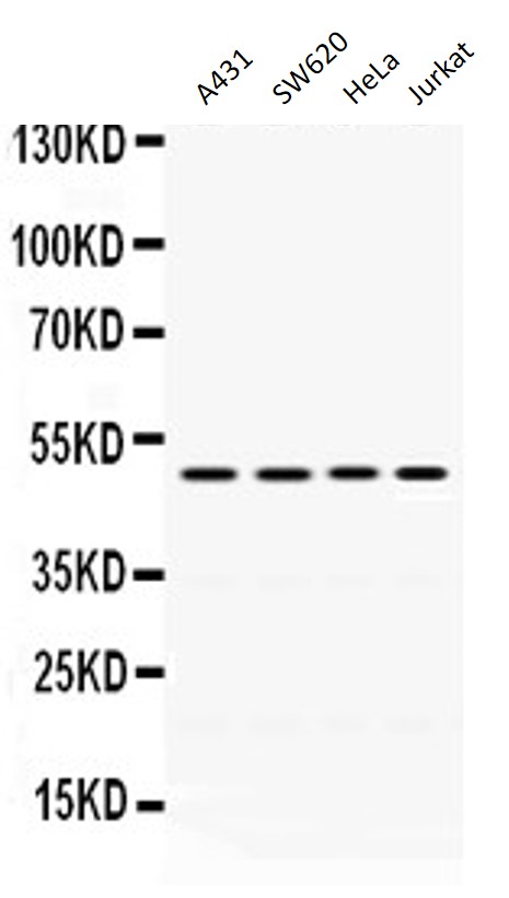
ARG58523 anti-DDB2 antibody WB image
Western blot: 40 µg of A431, 40 µg of SW620, 40 µg of HeLa and 40 µg of Jurkat cell lysates stained with ARG58523 anti-DDB2 antibody at 0.5 µg/ml dilution.
-

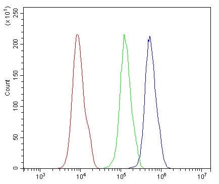
ARG58523 anti-DDB2 antibody FACS image
Flow Cytometry: 293T cells were blocked with 10% normal goat serum and then stained with ARG58523 anti-DDB2 antibody (blue) at 1 µg/10^6 cells for 30 min at 20°C, followed by incubation with DyLight®488 labelled secondary antibody. Isotype control antibody (green) was rabbit IgG (1 µg/10^6 cells) used under the same conditions. Unlabelled sample (red) was also used as a control.