ARG63154
anti-DDB1 antibody
anti-DDB1 antibody for IHC-Formalin-fixed paraffin-embedded sections,Western blot and Human,Mouse
Gene Regulation antibody
Overview
| Product Description | Goat Polyclonal antibody recognizes DDB1 |
|---|---|
| Tested Reactivity | Hu, Ms |
| Predict Reactivity | Cow, Rat, Dog |
| Tested Application | IHC-P, WB |
| Host | Goat |
| Clonality | Polyclonal |
| Isotype | IgG |
| Target Name | DDB1 |
| Antigen Species | Human |
| Immunogen | C-DLIKVVEELTRIH |
| Conjugation | Un-conjugated |
| Alternate Names | XPCE; XPCe; DDB p127 subunit; DDBa; UV-damaged DNA-binding factor; HBV X-associated protein 1; DDBA; UV-damaged DNA-binding protein 1; XPE; XAP-1; Damage-specific DNA-binding protein 1; XPE-BF; DNA damage-binding protein a; UV-DDB1; UV-DDB 1; XAP1; Xeroderma pigmentosum group E-complementing protein; XPE-binding factor; DNA damage-binding protein 1 |
Application Instructions
| Application Suggestion |
|
||||||
|---|---|---|---|---|---|---|---|
| Application Note | WB: Recommend incubate at RT for 1h. IHC-P: Antigen Retrieval: Steam tissue section in Citrate buffer (pH 6.0). * The dilutions indicate recommended starting dilutions and the optimal dilutions or concentrations should be determined by the scientist. |
Properties
| Form | Liquid |
|---|---|
| Purification | Purified from goat serum by antigen affinity chromatography. |
| Buffer | Tris saline (pH 7.3), 0.02% Sodium azide and 0.5% BSA. |
| Preservative | 0.02% Sodium azide |
| Stabilizer | 0.5% BSA |
| Concentration | 0.5 mg/ml |
| Storage Instruction | For continuous use, store undiluted antibody at 2-8°C for up to a week. For long-term storage, aliquot and store at -20°C or below. Storage in frost free freezers is not recommended. Avoid repeated freeze/thaw cycles. Suggest spin the vial prior to opening. The antibody solution should be gently mixed before use. |
| Note | For laboratory research only, not for drug, diagnostic or other use. |
Bioinformation
| Database Links | |
|---|---|
| Background | The protein encoded by this gene is the large subunit (p127) of the heterodimeric DNA damage-binding (DDB) complex while another protein (p48) forms the small subunit. This protein complex functions in nucleotide-excision repair and binds to DNA following UV damage. Defective activity of this complex causes the repair defect in patients with xeroderma pigmentosum complementation group E (XPE) - an autosomal recessive disorder characterized by photosensitivity and early onset of carcinomas. However, it remains for mutation analysis to demonstrate whether the defect in XPE patients is in this gene or the gene encoding the small subunit. In addition, Best vitelliform mascular dystrophy is mapped to the same region as this gene on 11q, but no sequence alternations of this gene are demonstrated in Best disease patients. The protein encoded by this gene also functions as an adaptor molecule for the cullin 4 (CUL4) ubiquitin E3 ligase complex by facilitating the binding of substrates to this complex and the ubiquitination of proteins. [provided by RefSeq, May 2012] |
| Research Area | Gene Regulation antibody |
| Calculated MW | 127 kDa |
| PTM | Phosphorylated by ABL1. Ubiquitinated by CUL4A. Subsequently degraded by ubiquitin-dependent proteolysis. |
Images (4) Click the Picture to Zoom In
-
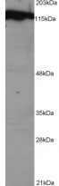
ARG63154 anti-DDB1 antibody WB image
Western Blot: NSO lysate (1E5 cells per lane) stained with ARG63154 anti-DDB1 antibody at 1 µg/ml dilution.
-

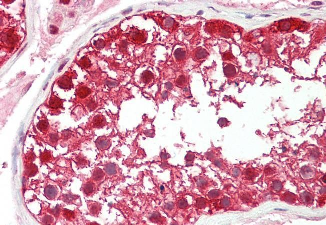
ARG63154 anti-DDB1 antibody IHC-P image
Immunohistochemistry: Paraffin-embedded Human testis tissue. Antigen Retrieval: Steam tissue section in Citrate buffer (pH 6.0). The tissue section was stained with ARG63154 anti-DDB1 antibody at 5 µg/ml dilution followed by AP-staining.
-

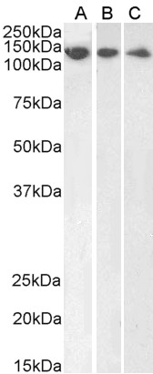
ARG63154 anti-DDB1 antibody WB image
Western blot: 35 µg of HeLa (A), HepG2 (B) and Jurkat (C) cell lysates (in RIPA buffer) stained with ARG63154 anti-DDB1 antibody at 1 µg/ml dilution and incubated at RT for 1 hour.
-
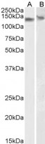
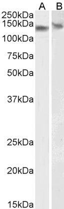
ARG63154 anti-DDB1 antibody WB image
Western blot: 35 µg of NIH/3T3 (A) and NSO (B) cell lysates (in RIPA buffer) stained with ARG63154 anti-DDB1 antibody at 0.01 µg/ml dilution and incubated at RT for 1 hour.