ARG64940
anti-DBP / Vitamin D binding protein antibody
anti-DBP / Vitamin D binding protein antibody for Western blot and Human
Cancer antibody; Metabolism antibody; Signaling Transduction antibody
Overview
| Product Description | Goat Polyclonal antibody recognizes DBP / Vitamin D binding protein |
|---|---|
| Tested Reactivity | Hu |
| Predict Reactivity | Ms, Rat, Dog |
| Tested Application | WB |
| Host | Goat |
| Clonality | Polyclonal |
| Isotype | IgG |
| Target Name | DBP / Vitamin D binding protein |
| Antigen Species | Human |
| Immunogen | ERGRDYEKNKVCK; the sequence is corresponding to internal sequence (near the N terminal) amino acids 18-30 of Human Vitamin D Binding protein (NP_000574.2). |
| Conjugation | Un-conjugated |
| Alternate Names | GRD3; DBP/GC; HEL-S-51; VDBG; VDB; Gc-globulin; DBP; VDBP; Vitamin D-binding protein; Group-specific component |
Application Instructions
| Application Suggestion |
|
||||
|---|---|---|---|---|---|
| Application Note | WB: Recommend incubate at RT for 1h. * The dilutions indicate recommended starting dilutions and the optimal dilutions or concentrations should be determined by the scientist. |
Properties
| Form | Liquid |
|---|---|
| Purification | Purified from goat serum by antigen affinity chromatography. |
| Buffer | Tris saline (pH 7.3), 0.02% Sodium azide and 0.5% BSA. |
| Preservative | 0.02% Sodium azide |
| Stabilizer | 0.5% BSA |
| Concentration | 0.5 mg/ml |
| Storage Instruction | For continuous use, store undiluted antibody at 2-8°C for up to a week. For long-term storage, aliquot and store at -20°C or below. Storage in frost free freezers is not recommended. Avoid repeated freeze/thaw cycles. Suggest spin the vial prior to opening. The antibody solution should be gently mixed before use. |
| Note | For laboratory research only, not for drug, diagnostic or other use. |
Bioinformation
| Database Links | |
|---|---|
| Gene Full Name | group-specific component (vitamin D binding protein) |
| Background | The protein encoded by this gene belongs to the albumin gene family. It is a multifunctional protein found in plasma, ascitic fluid, cerebrospinal fluid and on the surface of many cell types. It binds to vitamin D and its plasma metabolites and transports them to target tissues. Alternatively spliced transcript variants encoding different isoforms have been found for this gene.[provided by RefSeq, Feb 2011] |
| Research Area | Cancer antibody; Metabolism antibody; Signaling Transduction antibody |
| Calculated MW | 53 kDa |
| PTM | Allele GC*1S is O-glycosylated at Thr-436 (PubMed:20079467). The trisaccharide sugar moiety can be modified by the successive removal of neuraminic acid and galactose leaving an O-linked N-acetyl-galactosamine. This conversion is thought to produce a macrophage-activating factor (Gc-MAF). Only a minor proportion of plasma GC is O-glycosylated (PubMed:17360250). The potential N-glycosylation site predicted at Asn-288 is thought to be nonglycosylated. |
Images (1) Click the Picture to Zoom In
-
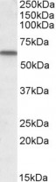
ARG64940 anti-DBP / Vitamin D binding protein antibody WB image
Western blot: Human Lung lysate (35 µg protein in RIPA buffer) stained with ARG64940 anti-DBP / Vitamin D binding protein antibody at 1 µg/ml dilution.


