ARG40921
anti-DBP / Vitamin D binding protein antibody
anti-DBP / Vitamin D binding protein antibody for IHC-Formalin-fixed paraffin-embedded sections,Western blot and Human,Mouse
Overview
| Product Description | Rabbit Polyclonal antibody recognizes DBP / Vitamin D binding protein |
|---|---|
| Tested Reactivity | Hu, Ms |
| Predict Reactivity | Rat |
| Tested Application | IHC-P, WB |
| Host | Rabbit |
| Clonality | Polyclonal |
| Isotype | IgG |
| Target Name | DBP / Vitamin D binding protein |
| Antigen Species | Human |
| Immunogen | Recombinant protein corresponding to L17-E256 of Human DBP. |
| Conjugation | Un-conjugated |
| Alternate Names | GRD3; DBP/GC; HEL-S-51; VDBG; VDB; Gc-globulin; DBP; VDBP; Vitamin D-binding protein; Group-specific component |
Application Instructions
| Application Suggestion |
|
||||||
|---|---|---|---|---|---|---|---|
| Application Note | IHC-P: Antigen Retrieval: Heat mediation was performed in Citrate buffer (pH 6.0, epitope retrieval solution) for 20 min. * The dilutions indicate recommended starting dilutions and the optimal dilutions or concentrations should be determined by the scientist. |
Properties
| Form | Liquid |
|---|---|
| Purification | Affinity purification with immunogen. |
| Buffer | 0.2% Na2HPO4, 0.9% NaCl, 0.05% Sodium azide and 5% BSA. |
| Preservative | 0.05% Sodium azide |
| Stabilizer | 5% BSA |
| Concentration | 0.5 mg/ml |
| Storage Instruction | For continuous use, store undiluted antibody at 2-8°C for up to a week. For long-term storage, aliquot and store at -20°C or below. Storage in frost free freezers is not recommended. Avoid repeated freeze/thaw cycles. Suggest spin the vial prior to opening. The antibody solution should be gently mixed before use. |
| Note | For laboratory research only, not for drug, diagnostic or other use. |
Bioinformation
| Database Links | |
|---|---|
| Gene Symbol | GC |
| Gene Full Name | group-specific component (vitamin D binding protein) |
| Background | The protein encoded by this gene belongs to the albumin gene family. It is a multifunctional protein found in plasma, ascitic fluid, cerebrospinal fluid and on the surface of many cell types. It binds to vitamin D and its plasma metabolites and transports them to target tissues. Alternatively spliced transcript variants encoding different isoforms have been found for this gene.[provided by RefSeq, Feb 2011] |
| Function | Multifunctional protein found in plasma, ascitic fluid, cerebrospinal fluid, and urine and on the surface of many cell types. In plasma, it carries the vitamin D sterols and prevents polymerization of actin by binding its monomers. DBP associates with membrane-bound immunoglobulin on the surface of B-lymphocytes and with IgG Fc receptor on the membranes of T-lymphocytes. [UniProt] |
| Cellular Localization | Secreted. [UniProt] |
| Calculated MW | 53 kDa |
| PTM | Allele GC*1S is O-glycosylated at Thr-436 (PubMed:20079467). The trisaccharide sugar moiety can be modified by the successive removal of neuraminic acid and galactose leaving an O-linked N-acetyl-galactosamine. This conversion is thought to produce a macrophage-activating factor (Gc-MAF). Only a minor proportion of plasma GC is O-glycosylated (PubMed:17360250). The potential N-glycosylation site predicted at Asn-288 is thought to be nonglycosylated. [UniProt] |
Images (5) Click the Picture to Zoom In
-
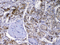
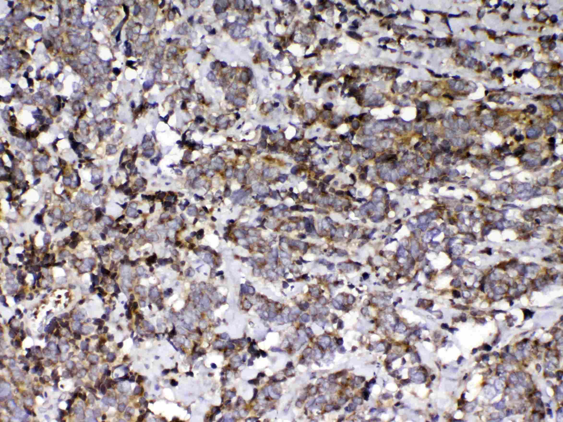
ARG40921 anti-DBP / Vitamin D binding protein antibody IHC-P image
Immunohistochemistry: Paraffin-embedded Human liver cancer tissue. Antigen Retrieval: Heat mediation was performed in Citrate buffer (pH 6.0, epitope retrieval solution) for 20 min. The tissue section was blocked with 10% goat serum. The tissue section was then stained with ARG40921 anti-DBP / Vitamin D binding protein antibody at 1 µg/ml dilution, overnight at 4°C.
-

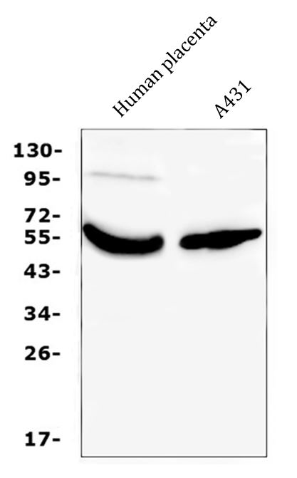
ARG40921 anti-DBP / Vitamin D binding protein antibody WB image
Western blot: 50 µg of samples under reducing conditions. Human placenta and A431 whole cell lysates stained with ARG40921 anti-DBP / Vitamin D binding protein antibody at 0.5 µg/ml, overnight at 4°C.
-
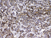
ARG40921 anti-DBP / Vitamin D binding protein antibody IHC-P image
Immunohistochemistry: Paraffin-embedded Human lung cancer tissue. Antigen Retrieval: Heat mediation was performed in Citrate buffer (pH 6.0, epitope retrieval solution) for 20 min. The tissue section was blocked with 10% goat serum. The tissue section was then stained with ARG40921 anti-DBP / Vitamin D binding protein antibody at 1 µg/ml dilution, overnight at 4°C.
-
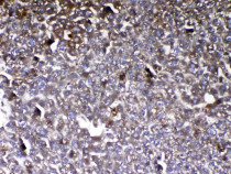
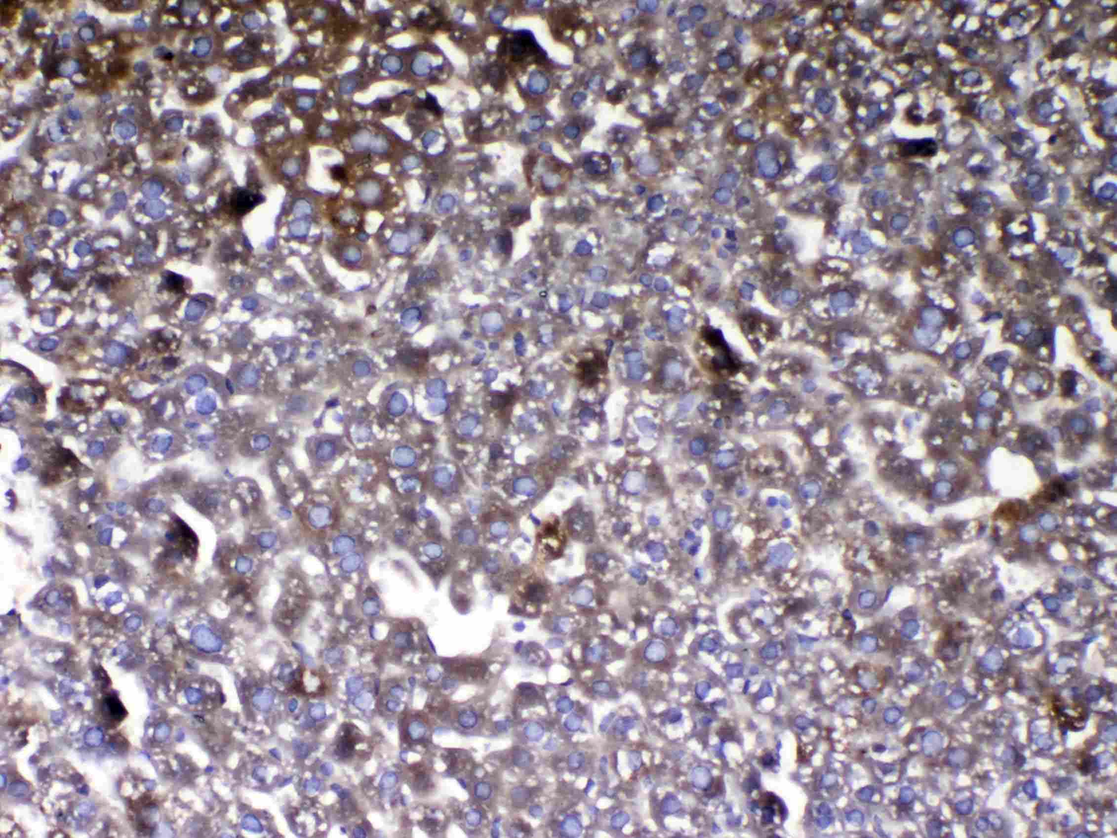
ARG40921 anti-DBP / Vitamin D binding protein antibody IHC-P image
Immunohistochemistry: Paraffin-embedded Mouse liver tissue. Antigen Retrieval: Heat mediation was performed in Citrate buffer (pH 6.0, epitope retrieval solution) for 20 min. The tissue section was blocked with 10% goat serum. The tissue section was then stained with ARG40921 anti-DBP / Vitamin D binding protein antibody at 1 µg/ml dilution, overnight at 4°C.
-
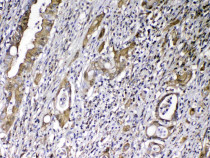
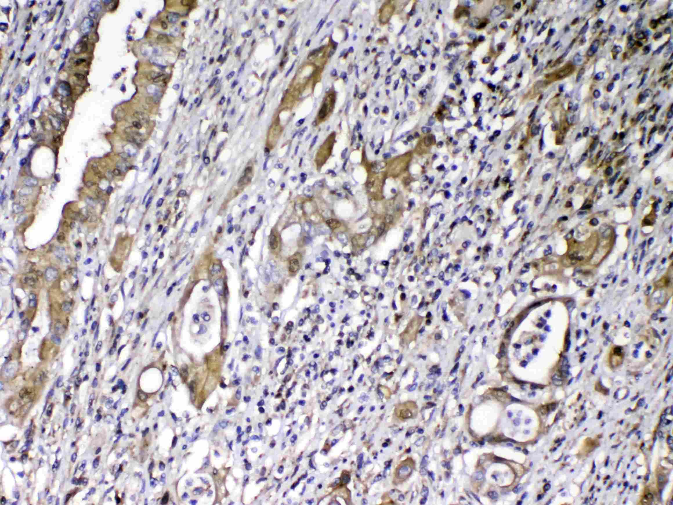
ARG40921 anti-DBP / Vitamin D binding protein antibody IHC-P image
Immunohistochemistry: Paraffin-embedded Human rectal cancer tissue. Antigen Retrieval: Heat mediation was performed in Citrate buffer (pH 6.0, epitope retrieval solution) for 20 min. The tissue section was blocked with 10% goat serum. The tissue section was then stained with ARG40921 anti-DBP / Vitamin D binding protein antibody at 1 µg/ml dilution, overnight at 4°C.