ARG58506
anti-DBI antibody
anti-DBI antibody for Flow cytometry,ICC/IF,IHC-Formalin-fixed paraffin-embedded sections,Western blot and Human
Overview
| Product Description | Rabbit Polyclonal antibody recognizes DBI |
|---|---|
| Tested Reactivity | Hu |
| Tested Application | FACS, ICC/IF, IHC-P, WB |
| Host | Rabbit |
| Clonality | Polyclonal |
| Isotype | IgG |
| Target Name | DBI |
| Antigen Species | Human |
| Immunogen | E. coli-derived Human DBI recombinant protein (Position: S2-I87). Human DBI shares 77.9% amino acid (aa) sequence identity with both Mouse and Rat DBI. |
| Conjugation | Un-conjugated |
| Alternate Names | DBI; ACBP; Acyl-CoA-binding protein; Endozepine; ACBD1; Diazepam-binding inhibitor; CCK-RP; EP |
Application Instructions
| Application Suggestion |
|
||||||||||
|---|---|---|---|---|---|---|---|---|---|---|---|
| Application Note | IHC-P: Antigen Retrieval: Heat mediation was performed in Citrate buffer (pH 6.0) for 20 min. * The dilutions indicate recommended starting dilutions and the optimal dilutions or concentrations should be determined by the scientist. |
Properties
| Form | Liquid |
|---|---|
| Purification | Affinity purification with immunogen. |
| Buffer | 0.9% NaCl, 0.2% Na2HPO4, 0.05% Sodium azide and 5% BSA. |
| Preservative | 0.05% Sodium azide |
| Stabilizer | 5% BSA |
| Concentration | 0.5 mg/ml |
| Storage Instruction | For continuous use, store undiluted antibody at 2-8°C for up to a week. For long-term storage, aliquot and store at -20°C or below. Storage in frost free freezers is not recommended. Avoid repeated freeze/thaw cycles. Suggest spin the vial prior to opening. The antibody solution should be gently mixed before use. |
| Note | For laboratory research only, not for drug, diagnostic or other use. |
Bioinformation
| Database Links | |
|---|---|
| Gene Symbol | DBI |
| Gene Full Name | diazepam binding inhibitor (GABA receptor modulator, acyl-CoA binding protein) |
| Background | This gene encodes diazepam binding inhibitor, a protein that is regulated by hormones and is involved in lipid metabolism and the displacement of beta-carbolines and benzodiazepines, which modulate signal transduction at type A gamma-aminobutyric acid receptors located in brain synapses. The protein is conserved from yeast to mammals, with the most highly conserved domain consisting of seven contiguous residues that constitute the hydrophobic binding site for medium- and long-chain acyl-Coenzyme A esters. Diazepam binding inhibitor is also known to mediate the feedback regulation of pancreatic secretion and the postprandial release of cholecystokinin, in addition to its role as a mediator in corticotropin-dependent adrenal steroidogenesis. Three pseudogenes located on chromosomes 6, 8 and 16 have been identified. Multiple transcript variants encoding different isoforms have been described for this gene. [provided by RefSeq, Jul 2008] |
| Function | Binds medium- and long-chain acyl-CoA esters with very high affinity and may function as an intracellular carrier of acyl-CoA esters. It is also able to displace diazepam from the benzodiazepine (BZD) recognition site located on the GABA type A receptor. It is therefore possible that this protein also acts as a neuropeptide to modulate the action of the GABA receptor. [UniProt] |
| Cellular Localization | Endoplasmic reticulum. Golgi apparatus. Golgi localization is dependent on ligand binding (PubMed:17953517). [UniProt] |
| Calculated MW | 10 kDa |
Images (7) Click the Picture to Zoom In
-
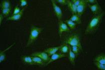
ARG58506 anti-DBI antibody ICC/IF image
Immunofluorescence: U2OS cells were blocked with 10% goat serum and then stained with ARG58506 anti-DBI antibody (green) at 2 µg/ml dilution, overnight at 4°C. DAPI (blue) for nuclear staining.
-
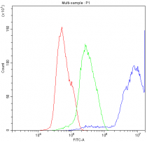
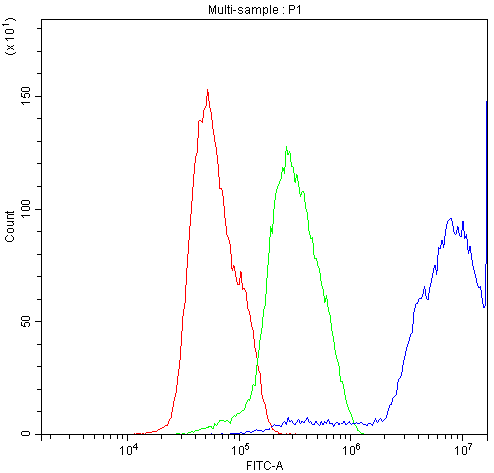
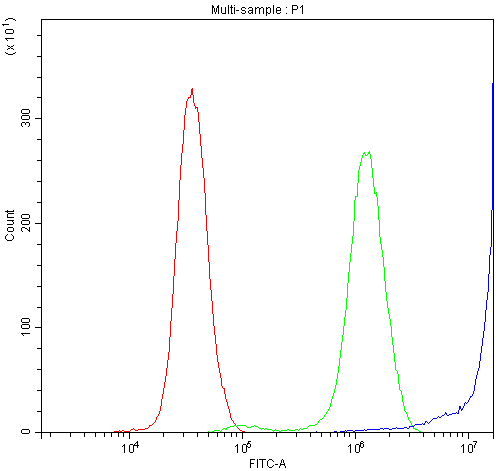
ARG58506 anti-DBI antibody FACS image
Flow Cytometry: MCF-7 cells were blocked with 10% normal goat serum, and then stained with ARG58506 anti-DBI antibody (blue) at 1 µg/10^6 cells for 30 min at 20°C, followed by DyLight®488 labelled secondary antibody. Isotype control antibody (green) was rabbit IgG (1 µg/10^6 cells) used under the same conditions. Unlabelled sample (red) was also used as a control.
-
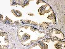
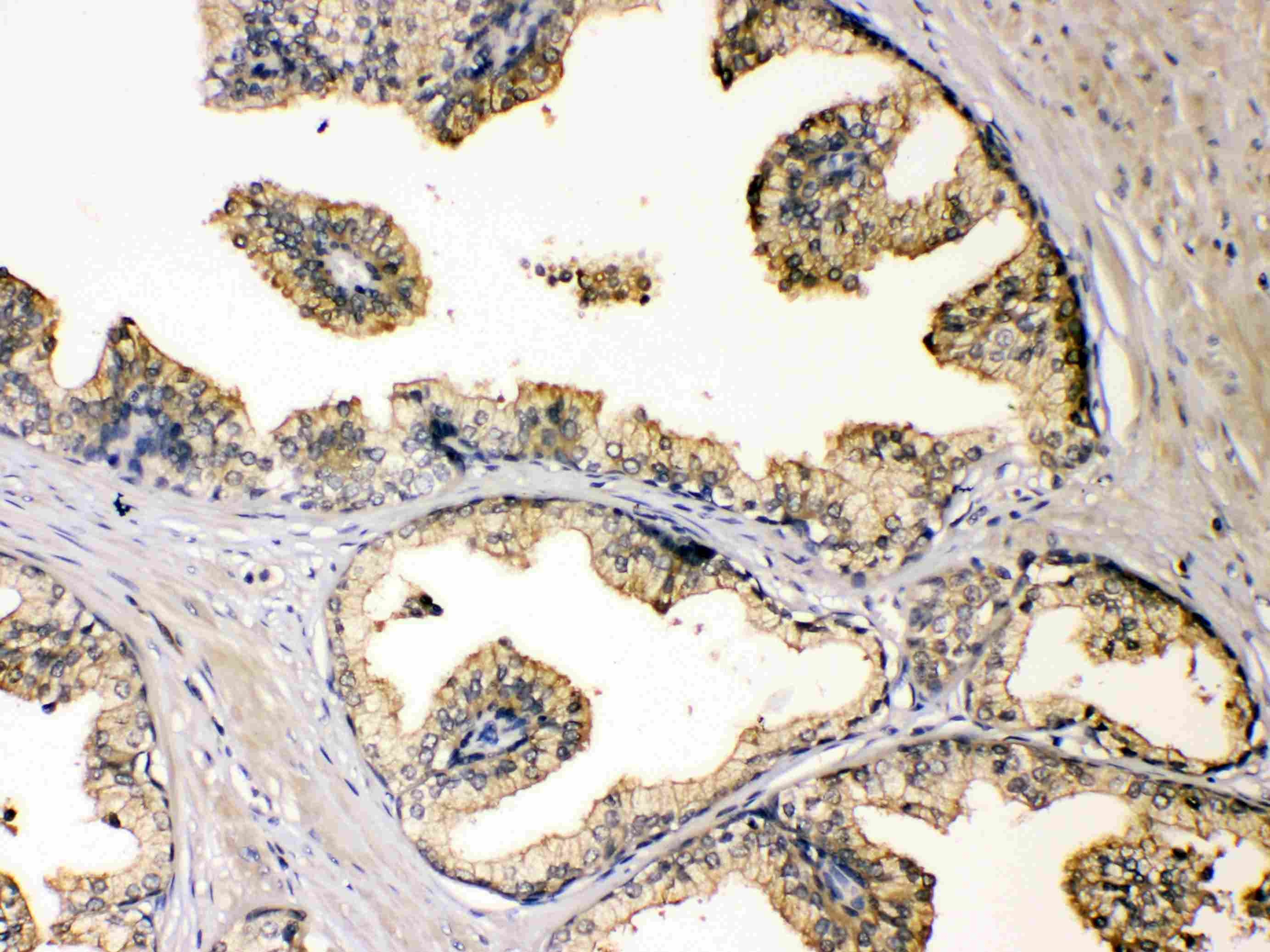
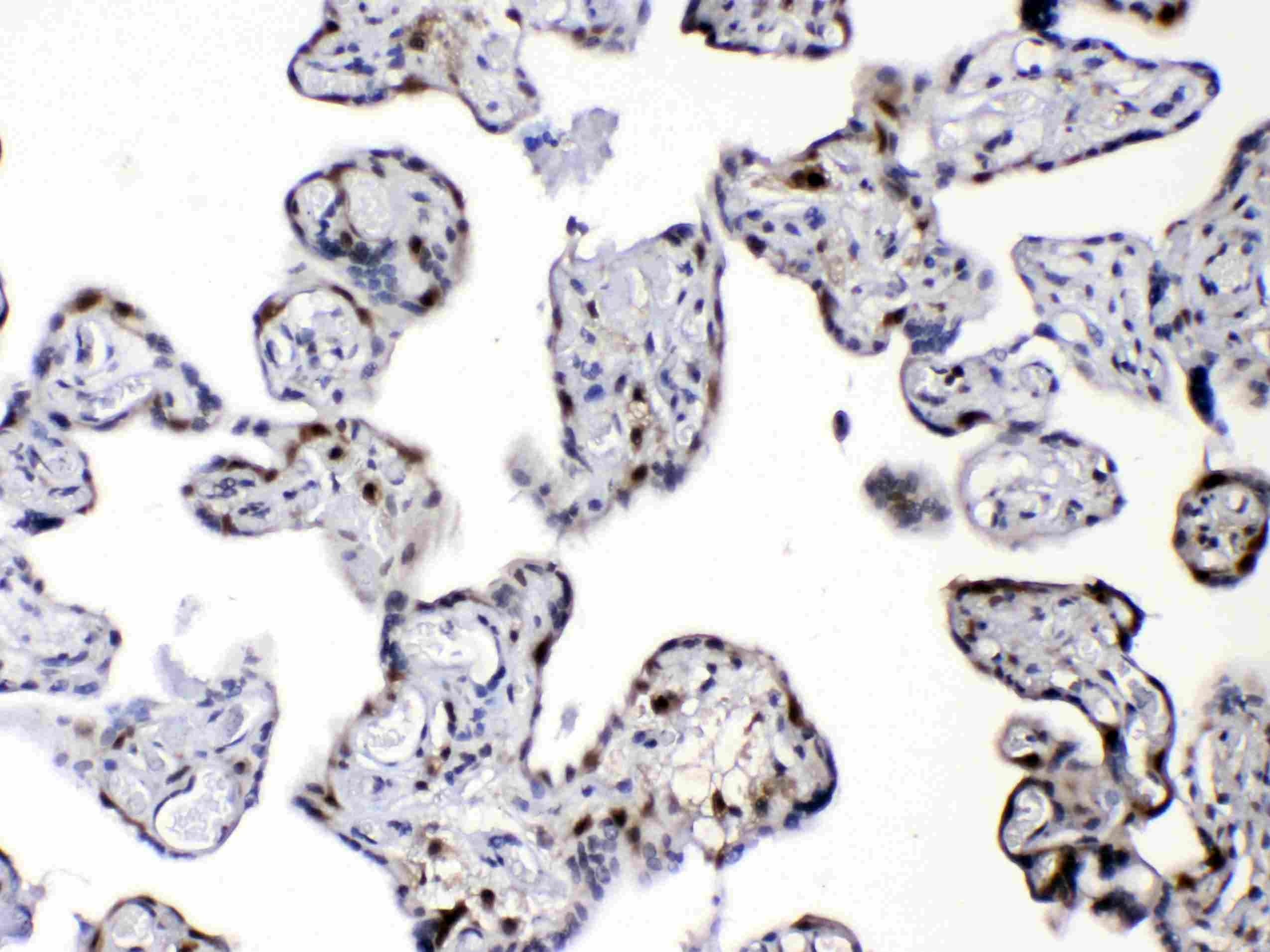
ARG58506 anti-DBI antibody IHC-P image
Immunohistochemistry: Paraffin-embedded Human prostatic cancer tissue. Antigen Retrieval: Heat mediated was performed in Citrate buffer (pH 6.0, epitope retrieval solution) for 20 min. The tissue section was blocked with 10% goat serum. The tissue section was then stained with ARG58506 anti-DBI antibody at 1 µg/ml dilution, overnight at 4°C.
-

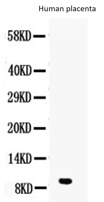
ARG58506 anti-DBI antibody WB image
Western blot: 50 µg of sample under reducing conditions. Human placenta lysate stained with ARG58506 anti-DBI antibody at 0.5 µg/ml, overnight at 4°C.
-
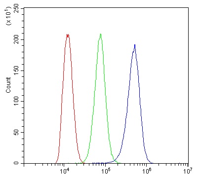
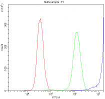
ARG58506 anti-DBI antibody FACS image
Flow Cytometry: THP-1 cells were blocked with 10% normal goat serum and then stained with ARG58506 anti-DBI antibody (blue) at 1 µg/10^6 cells for 30 min at 20°C, followed by incubation with DyLight®488 labelled secondary antibody. Isotype control antibody (green) was rabbit IgG (1 µg/10^6 cells) used under the same conditions. Unlabelled sample (red) was also used as a control.
-
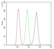
ARG58506 anti-DBI antibody FACS image
Flow Cytometry: PC-3 cells were blocked with 10% normal goat serum, and then stained with ARG58506 anti-DBI antibody (blue) at 1 µg/10^6 cells for 30 min at 20°C, followed by DyLight®488 labelled secondary antibody. Isotype control antibody (green) was rabbit IgG (1 µg/10^6 cells) used under the same conditions. Unlabelled sample (red) was also used as a control.
-
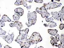
ARG58506 anti-DBI antibody IHC-P image
Immunohistochemistry: Paraffin-embedded Human placentas. Antigen Retrieval: Heat mediated was performed in Citrate buffer (pH 6.0, epitope retrieval solution) for 20 min. The tissue section was blocked with 10% goat serum. The tissue section was then stained with ARG58506 anti-DBI antibody at 1 µg/ml dilution, overnight at 4°C.