ARG53215
anti-Cytokeratin 8 antibody
anti-Cytokeratin 8 antibody for IHC-Formalin-fixed paraffin-embedded sections,Western blot and Human
Cancer antibody; Signaling Transduction antibody

1
Overview
| Product Description | Rabbit Polyclonal antibody recognizes Cytokeratin 8 |
|---|---|
| Tested Reactivity | Hu |
| Tested Application | IHC-P, WB |
| Host | Rabbit |
| Clonality | Polyclonal |
| Isotype | IgG |
| Target Name | Cytokeratin 8 |
| Antigen Species | Human |
| Immunogen | Recombinant protein corresponding to aa. 1-483 of Human Cytokeratin 8. |
| Conjugation | Un-conjugated |
| Alternate Names | Keratin, type II cytoskeletal 8; KO; CYK8; CK-8; Type-II keratin Kb8; K2C8; CARD2; Keratin-8; K8; CK8; Cytokeratin-8 |
Application Instructions
| Application Suggestion |
|
||||||
|---|---|---|---|---|---|---|---|
| Application Note | IHC-P: Antigen Retrieval: Boil tissue section in 10mM citrate buffer, pH 6.0 for 10 min followed by cooling at RT for 20 min. Incubation Time: 10 min at RT. * The dilutions indicate recommended starting dilutions and the optimal dilutions or concentrations should be determined by the scientist. |
||||||
| Positive Control | Breast Carcinoma, Lung, Skin, HeLa Cell Lysate |
Properties
| Form | Liquid |
|---|---|
| Purification | Immunogen affinity purified |
| Buffer | PBS (pH 7.6), 1% BSA and < 0.1% Sodium azide |
| Preservative | < 0.1% Sodium azide |
| Stabilizer | 1% BSA |
| Storage Instruction | For continuous use, store undiluted antibody at 2-8°C for up to a week. For long-term storage, aliquot and store at -20°C or below. Storage in frost free freezers is not recommended. Avoid repeated freeze/thaw cycles. Suggest spin the vial prior to opening. The antibody solution should be gently mixed before use. |
| Note | For laboratory research only, not for drug, diagnostic or other use. |
Bioinformation
| Database Links | |
|---|---|
| Background | Keratin 8 belongs to the type B (basic) subfamily of high molecular weight keratins and exists in combination with keratin 18. Keratin 8 is primarily found in the non-squamous epithelia and is present in majority of adenocarcinomas and ductal carcinomas. It is absent in squamous cell carcinomas. Hepatocellular carcinomas are defined by the use of antibodies that recognize only cytokeratin polypeptides 8 and 18. |
| Cellular Localization | Cytoplasm, Membrane |
| Research Area | Cancer antibody; Signaling Transduction antibody |
| Calculated MW | 54 kDa |
| PTM | Phosphorylation on serine residues is enhanced during EGF stimulation and mitosis. Ser-74 phosphorylation plays an important role in keratin filament reorganization. O-glycosylated. O-GlcNAcylation at multiple sites increases solubility, and decreases stability by inducing proteasomal degradation. O-glycosylated (O-GlcNAcylated), in a cell cycle-dependent manner. |
Images (2) Click the Picture to Zoom In
-
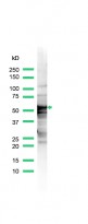
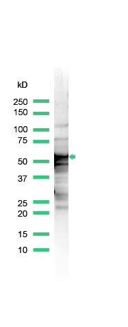
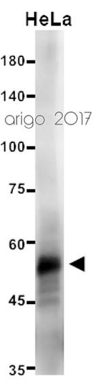
ARG53215 anti-Cytokeratin 8 antibody WB image
Western blot: HeLa cell lysate stained with ARG53215 anti-Cytokeratin 8 antibody.
-
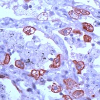
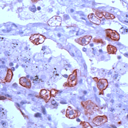
ARG53215 anti-Cytokeratin 8 antibody IHC-P image
Immunohistochemistry: Human lung stained with ARG53215 anti-Cytokeratin 8 antibody.
Customer's Feedback
Good
Review for anti-Cytokeratin 8 antibody
Application:WB
Sample:HeLa
Sample Loading Amount:20 µg
Primary Antibody Dilution Factor:1:25
Primary Antibody Incubation Time:overnight
Primary Antibody Incubation Temperature:4 ºC


