ARG66386
anti-Cytokeratin 6 antibody [SQab18113]
anti-Cytokeratin 6 antibody [SQab18113] for IHC-Formalin-fixed paraffin-embedded sections,Immunoprecipitation,Western blot and Human,Mouse,Rat
Overview
| Product Description | Recombinant Rabbit Monoclonal antibody [SQab18113] recognizes Cytokeratin 6 |
|---|---|
| Tested Reactivity | Hu, Ms, Rat |
| Tested Application | IHC-P, IP, WB |
| Host | Rabbit |
| Clonality | Monoclonal |
| Clone | SQab18113 |
| Isotype | IgG |
| Target Name | Cytokeratin 6 |
| Antigen Species | Human |
| Immunogen | Synthetic peptide around the C-terminus of Human Cytokeratin 6. |
| Conjugation | Un-conjugated |
| Alternate Names | CK-6A; K6D; Cytokeratin-6A; CK-6D; K6C; K6A; Type-II keratin Kb6; PC3; CK6D; KRT6D; CK6A; CK6C; allergen Hom s 5; Keratin, type II cytoskeletal 6A; Keratin-6A; Cytokeratin-6D; KRT6C |
Application Instructions
| Application Suggestion |
|
||||||||
|---|---|---|---|---|---|---|---|---|---|
| Application Note | IHC-P: Antigen Retrieval: Heat mediated was performed using Tris/EDTA buffer (pH 9.0). * The dilutions indicate recommended starting dilutions and the optimal dilutions or concentrations should be determined by the scientist. |
Properties
| Form | Liquid |
|---|---|
| Purification | Purification with Protein A. |
| Buffer | PBS, 0.01% Sodium azide, 40% Glycerol and 0.05% BSA. |
| Preservative | 0.01% Sodium azide |
| Stabilizer | 40% Glycerol and 0.05% BSA |
| Storage Instruction | For continuous use, store undiluted antibody at 2-8°C for up to a week. For long-term storage, aliquot and store at -20°C. Storage in frost free freezers is not recommended. Avoid repeated freeze/thaw cycles. Suggest spin the vial prior to opening. The antibody solution should be gently mixed before use. |
| Note | For laboratory research only, not for drug, diagnostic or other use. |
Bioinformation
| Database Links | |
|---|---|
| Gene Symbol | KRT6A |
| Gene Full Name | keratin 6A, type II |
| Background | The protein encoded by this gene is a member of the keratin gene family. The type II cytokeratins consist of basic or neutral proteins which are arranged in pairs of heterotypic keratin chains coexpressed during differentiation of simple and stratified epithelial tissues. As many as six of this type II cytokeratin (KRT6) have been identified; the multiplicity of the genes is attributed to successive gene duplication events. The genes are expressed with family members KRT16 and/or KRT17 in the filiform papillae of the tongue, the stratified epithelial lining of oral mucosa and esophagus, the outer root sheath of hair follicles, and the glandular epithelia. This KRT6 gene in particular encodes the most abundant isoform. Mutations in these genes have been associated with pachyonychia congenita. In addition, peptides from the C-terminal region of the protein have antimicrobial activity against bacterial pathogens. The type II cytokeratins are clustered in a region of chromosome 12q12-q13. [provided by RefSeq, Oct 2014] |
| Function | Epidermis-specific type I keratin involved in wound healing. Involved in the activation of follicular keratinocytes after wounding, while it does not play a major role in keratinocyte proliferation or migration. Participates in the regulation of epithelial migration by inhibiting the activity of SRC during wound repair. [UniProt] |
| Calculated MW | 60 kDa |
Images (4) Click the Picture to Zoom In
-
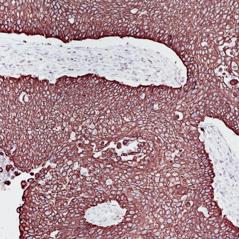
ARG66386 anti-Cytokeratin 6 antibody [SQab18113] IHC-P image
Immunohistochemistry: Formalin-fixed and paraffin-embedded Human esophageal cancer stained with ARG66386 anti-Cytokeratin 6 antibody [SQab18113] at 1:50 dilution. Antigen Retrieval: Heat mediated was performed using Tris/EDTA buffer (pH 9.0).
-
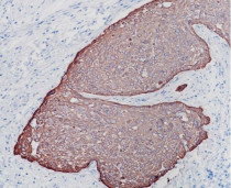
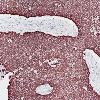
ARG66386 anti-Cytokeratin 6 antibody [SQab18113] IHC-P image
Immunohistochemistry: Formalin-fixed and paraffin-embedded Human cervix cancer stained with ARG66386 anti-Cytokeratin 6 antibody [SQab18113] at 1:50 dilution. Antigen Retrieval: Heat mediated was performed using Tris/EDTA buffer (pH 9.0).
-
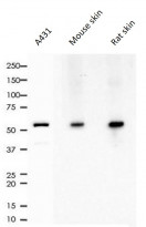
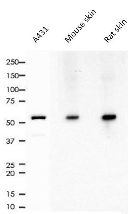
ARG66386 anti-Cytokeratin 6 antibody [SQab18113] WB image
Western blot: 10 µg of A431, Mouse skin and Rat skin lysates stained with ARG66386 anti-Cytokeratin 6 antibody [SQab18113] at 1:10000 dilution.
-

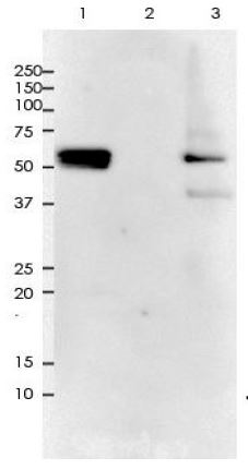
ARG66386 anti-Cytokeratin 6 antibody [SQab18113] IP image
Immunoprecipitation: 0.4 mg of A431 whole cell lysate was immunoprecipitated (1:50 dilution) and stained with ARG66386 anti-Cytokeratin 6 antibody [SQab18113].
Lane 1: Immunoprecipitation in A431 whole cell lysate
Lane 2: PBS instead of Primary Ab in A431 whole cell lysate
Lane 3: A431 whole cell lysate, 10 µg (input)