ARG66970
anti-Cytokeratin 5 + 6 antibody
anti-Cytokeratin 5 + 6 antibody for ICC/IF,IHC-Formalin-fixed paraffin-embedded sections,Western blot and Human
Overview
| Product Description | Rabbit Polyclonal antibody recognizes Cytokeratin 5 + 6 |
|---|---|
| Tested Reactivity | Hu |
| Tested Application | ICC/IF, IHC-P, WB |
| Host | Rabbit |
| Clonality | Polyclonal |
| Isotype | IgG |
| Target Name | Cytokeratin 5 + 6 |
| Antigen Species | Human |
| Immunogen | Synthetic peptide around the middle region of Human Cytokeratin 5. |
| Conjugation | Un-conjugated |
| Alternate Names | KRT5: DDD1; 58 kDa cytokeratin; Keratin, type II cytoskeletal 5; EBS2; Keratin-5; Cytokeratin-5; CK5; KRT5A; K5; CK-5; Type-II keratin Kb5; DDD KRT6A: CK-6A; K6D; Cytokeratin-6A; CK-6D; K6C; K6A; Type-II keratin Kb6; PC3; CK6D; KRT6D; CK6A; CK6C; allergen Hom s 5; Keratin, type II cytoskeletal 6A; Keratin-6A; Cytokeratin-6D; KRT6C |
Application Instructions
| Application Suggestion |
|
||||||||
|---|---|---|---|---|---|---|---|---|---|
| Application Note | * The dilutions indicate recommended starting dilutions and the optimal dilutions or concentrations should be determined by the scientist. | ||||||||
| Positive Control | MDA-MB-231; Colorectal carcinoma; Breast carcinoma | ||||||||
| Observed Size | ~ 45 - 55 kDa |
Properties
| Form | Liquid |
|---|---|
| Purification | Affinity purified. |
| Buffer | 100 mM Tris Glycine (pH 7.0), 0.025% ProClin 300 and 20% Glycerol. |
| Preservative | 0.025% ProClin 300 |
| Stabilizer | 20% Glycerol |
| Storage Instruction | For continuous use, store undiluted antibody at 2-8°C for up to a week. For long-term storage, aliquot and store at -20°C. Storage in frost free freezers is not recommended. Avoid repeated freeze/thaw cycles. Suggest spin the vial prior to opening. The antibody solution should be gently mixed before use. |
| Note | For laboratory research only, not for drug, diagnostic or other use. |
Bioinformation
| Gene Symbol | KRT5; KRT6A |
|---|---|
| Gene Full Name | keratin 5, type II; keratin 6A, type II |
| Background | KRT5: The protein encoded by this gene is a member of the keratin gene family. The type II cytokeratins consist of basic or neutral proteins which are arranged in pairs of heterotypic keratin chains coexpressed during differentiation of simple and stratified epithelial tissues. This type II cytokeratin is specifically expressed in the basal layer of the epidermis with family member KRT14. Mutations in these genes have been associated with a complex of diseases termed epidermolysis bullosa simplex. The type II cytokeratins are clustered in a region of chromosome 12q12-q13. [provided by RefSeq, Jul 2008] KRT6A: The protein encoded by this gene is a member of the keratin gene family. The type II cytokeratins consist of basic or neutral proteins which are arranged in pairs of heterotypic keratin chains coexpressed during differentiation of simple and stratified epithelial tissues. As many as six of this type II cytokeratin (KRT6) have been identified; the multiplicity of the genes is attributed to successive gene duplication events. The genes are expressed with family members KRT16 and/or KRT17 in the filiform papillae of the tongue, the stratified epithelial lining of oral mucosa and esophagus, the outer root sheath of hair follicles, and the glandular epithelia. This KRT6 gene in particular encodes the most abundant isoform. Mutations in these genes have been associated with pachyonychia congenita. In addition, peptides from the C-terminal region of the protein have antimicrobial activity against bacterial pathogens. The type II cytokeratins are clustered in a region of chromosome 12q12-q13. [provided by RefSeq, Oct 2014] |
| Function | KRT6: Epidermis-specific type I keratin involved in wound healing. Involved in the activation of follicular keratinocytes after wounding, while it does not play a major role in keratinocyte proliferation or migration. Participates in the regulation of epithelial migration by inhibiting the activity of SRC during wound repair. [UniProt] |
| Calculated MW | 62 kDa |
Images (4) Click the Picture to Zoom In
-

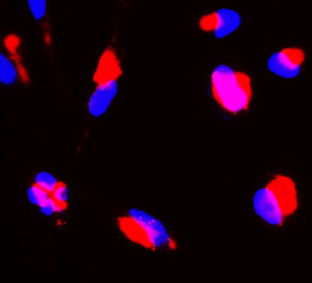
ARG66970 anti-Cytokeratin 5 + 6 antibody ICC/IF image
Immunofluorescence: MDA-MB-231 cells were fixed with 4% paraformaldehyde at RT for 10 min, permeabilized with 0.1% NP-40 at RT for 10 min and then cells were blocked with 5% BSA at RT for 30 min and stained with ARG66970 anti-Cytokeratin 5 + 6 antibody (red) at 1:150 dilution at 4°C for overnight. DAPI (blue) was used for nuclear staining.
-
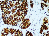
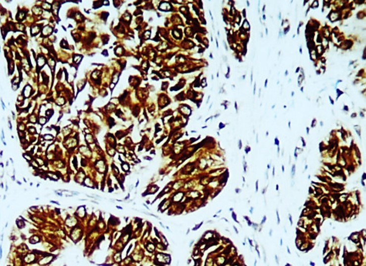
ARG66970 anti-Cytokeratin 5 + 6 antibody IHC-P image
Immunohistochemistry: Paraffin-embedded Human breast carcinoma tissue stained with ARG66970 anti-Cytokeratin 5 + 6 antibody at 1:100 dilution.
-
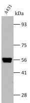
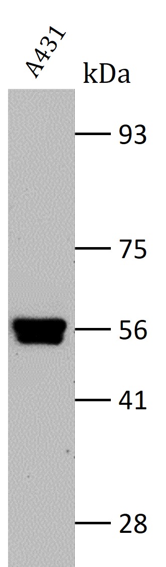
ARG66970 anti-Cytokeratin 5 + 6 antibody WB image
Western blot: A431 cell lysate stained with ARG66970 anti-Cytokeratin 5 + 6 antibody at 1:2000 dilution, overnight at 4°C.
-
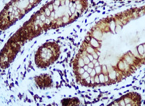
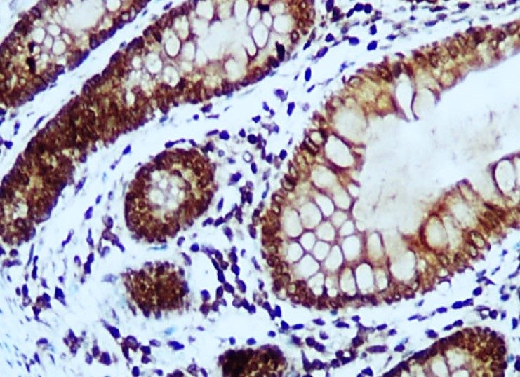
ARG66970 anti-Cytokeratin 5 + 6 antibody IHC-P image
Immunohistochemistry: Paraffin-embedded Human colorectal carcinoma tissue stained with ARG66970 anti-Cytokeratin 5 + 6 antibody at 1:100 dilution.