ARG53929
anti-Cytokeratin 19 antibody [A53-B/A2] (Biotin)
anti-Cytokeratin 19 antibody [A53-B/A2] (Biotin) for ELISA,Flow cytometry,ICC/IF,IHC-Formalin-fixed paraffin-embedded sections,Immunoprecipitation,Western blot and Human
Signaling Transduction antibody
Overview
| Product Description | Biotin-conjugated Mouse Monoclonal antibody [A53-B/A2] recognizes Cytokeratin 19 |
|---|---|
| Tested Reactivity | Hu |
| Tested Application | ELISA, FACS, ICC/IF, IHC-P, IP, WB |
| Specificity | The clone A53-B/A2 reacts with Rod domain of cytokeratin 19 (40 kDa) in human tissue. Cytokeratin 19 is not expressed in hepatocytes; it is often co-expressed with cytokeratin 7. |
| Host | Mouse |
| Clonality | Monoclonal |
| Clone | A53-B/A2 |
| Isotype | IgG2a |
| Target Name | Cytokeratin 19 |
| Antigen Species | Human |
| Immunogen | MCF-7 human breast adenocarcinoma cell line |
| Conjugation | Biotin |
| Alternate Names | Cytokeratin-19; K19; CK-19; K1CS; Keratin-19; CK19; Keratin, type I cytoskeletal 19 |
Application Instructions
| Application Suggestion |
|
||||||||||||||
|---|---|---|---|---|---|---|---|---|---|---|---|---|---|---|---|
| Application Note | * The dilutions indicate recommended starting dilutions and the optimal dilutions or concentrations should be determined by the scientist. | ||||||||||||||
| Positive Control | HT-29 | ||||||||||||||
| Observed Size | ~ 40 kDa |
Properties
| Form | Liquid |
|---|---|
| Purification Note | The purified antibody is conjugated with Biotin-LC-NHS under optimum conditions. The reagent is free of unconjugated biotin. |
| Buffer | PBS (pH 7.4) and 15 mM Sodium azide |
| Preservative | 15 mM Sodium azide |
| Concentration | 1 mg/ml |
| Storage Instruction | Aliquot and store in the dark at 2-8°C. Keep protected from prolonged exposure to light. Avoid repeated freeze/thaw cycles. Suggest spin the vial prior to opening. The antibody solution should be gently mixed before use. |
| Note | For laboratory research only, not for drug, diagnostic or other use. |
Bioinformation
| Database Links | |
|---|---|
| Gene Symbol | KRT19 |
| Gene Full Name | keratin 19, type I |
| Background | Cytokeratin 19 is a member of the keratin family. The keratins are intermediate filament proteins responsible for the structural integrity of epithelial cells and are subdivided into cytokeratins and hair keratins. The type I cytokeratins consist of acidic proteins which are arranged in pairs of heterotypic keratin chains. Unlike its related family members, this smallest known acidic cytokeratin is not paired with a basic cytokeratin in epithelial cells. It is specifically expressed in the periderm, the transiently superficial layer that envelopes the developing epidermis. The type I cytokeratins are clustered in a region of chromosome 17q12-q21. [provided by RefSeq, Jul 2008] |
| Function | Cytokeratin 19 involved in the organization of myofibers. Together with KRT8, helps to link the contractile apparatus to dystrophin at the costameres of striated muscle. [UniProt] |
| Highlight | Related products: Cytokeratin 19 antibodies; Anti-Mouse IgG secondary antibodies; Related news: Therapeutic strategies against PDAC |
| Research Area | Signaling Transduction antibody |
| Calculated MW | 44 kDa |
Images (2) Click the Picture to Zoom In
-
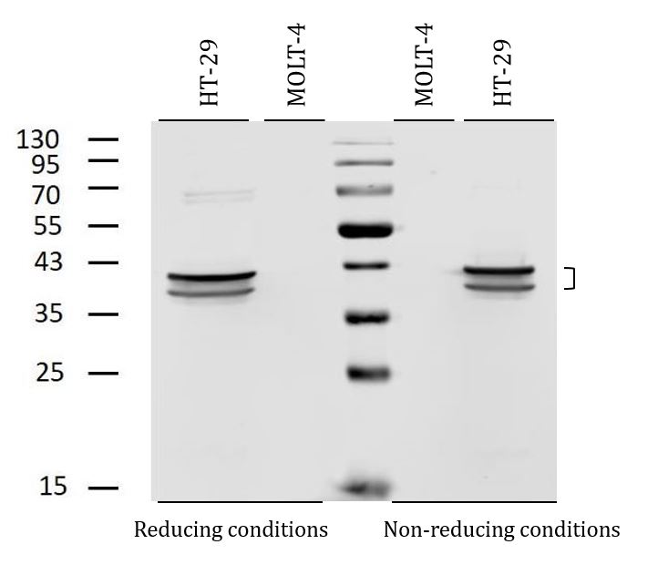
ARG53929 anti-Cytokeratin 19 antibody [A53-B/A2] (Biotin) WB image
Western blot: HT-29 (positive) and MOLT-4 (negative control) cell lysates stained with ARG53929 anti-Cytokeratin 19 antibody [A53-B/A2] (Biotin) at 2 µg/ml dilution, under reducing and non-reducing conditions.
-
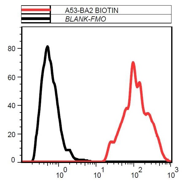
ARG53929 anti-Cytokeratin 19 antibody [A53-B/A2] (Biotin) FACS image
Flow Cytometry: MCF7 cells stained with ARG53929 anti-Cytokeratin 19 antibody [A53-B/A2] (Biotin).