ARG40920
anti-Cytokeratin 18 antibody
anti-Cytokeratin 18 antibody for Flow cytometry,IHC-Formalin-fixed paraffin-embedded sections,Western blot and Human,Mouse,Rat
Overview
| Product Description | Rabbit Polyclonal antibody recognizes Cytokeratin 18 |
|---|---|
| Tested Reactivity | Hu, Ms, Rat |
| Tested Application | FACS, IHC-P, WB |
| Host | Rabbit |
| Clonality | Polyclonal |
| Isotype | IgG |
| Target Name | Cytokeratin 18 |
| Antigen Species | Human |
| Immunogen | Recombinant protein corresponding to E204-H430 of Human Cytokeratin 18. |
| Conjugation | Un-conjugated |
| Alternate Names | Keratin, type I cytoskeletal 18; Cytokeratin-18; K18; CK-18; Cell proliferation-inducing gene 46 protein; Keratin-18; CYK18 |
Application Instructions
| Application Suggestion |
|
||||||||
|---|---|---|---|---|---|---|---|---|---|
| Application Note | IHC-P: Antigen Retrieval: Heat mediation was performed in Citrate buffer (pH 6.0, epitope retrieval solution) for 20 min. * The dilutions indicate recommended starting dilutions and the optimal dilutions or concentrations should be determined by the scientist. |
Properties
| Form | Liquid |
|---|---|
| Purification | Affinity purification with immunogen. |
| Buffer | 0.2% Na2HPO4, 0.9% NaCl, 0.05% Sodium azide and 5% BSA. |
| Preservative | 0.05% Sodium azide |
| Stabilizer | 5% BSA |
| Concentration | 0.5 mg/ml |
| Storage Instruction | For continuous use, store undiluted antibody at 2-8°C for up to a week. For long-term storage, aliquot and store at -20°C or below. Storage in frost free freezers is not recommended. Avoid repeated freeze/thaw cycles. Suggest spin the vial prior to opening. The antibody solution should be gently mixed before use. |
| Note | For laboratory research only, not for drug, diagnostic or other use. |
Bioinformation
| Database Links | |
|---|---|
| Gene Symbol | KRT18 |
| Gene Full Name | keratin 18, type I |
| Background | Cytokeratin 18, together with its filament partner Cytokeratin 8, are perhaps the most commonly found members of the intermediate filament gene family. They are expressed in single layer epithelial tissues of the body. Mutations in this gene have been linked to cryptogenic cirrhosis. Two transcript variants encoding the same protein have been found for this gene. [provided by RefSeq, Jul 2008] |
| Function | Cytokeratin 18 involved in the uptake of thrombin-antithrombin complexes by hepatic cells. When phosphorylated, plays a role in filament reorganization. Involved in the delivery of mutated CFTR to the plasma membrane. Together with KRT8, is involved in interleukin-6 (IL-6)-mediated barrier protection. [UniProt] |
| Cellular Localization | Cytoplasm, perinuclear region. Nucleus, nucleolus. [UniProt] |
| Calculated MW | 48 kDa |
| PTM | Phosphorylation at Ser-34 increases during mitosis. Hyperphosphorylated at Ser-53 in diseased cirrhosis liver. Phosphorylation increases by IL-6. Proteolytically cleaved by caspases during epithelial cell apoptosis. Cleavage occurs at Asp-238 by either caspase-3, caspase-6 or caspase-7. O-GlcNAcylation increases solubility, and decreases stability by inducing proteasomal degradation. [UniProt] |
Images (11) Click the Picture to Zoom In
-
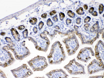
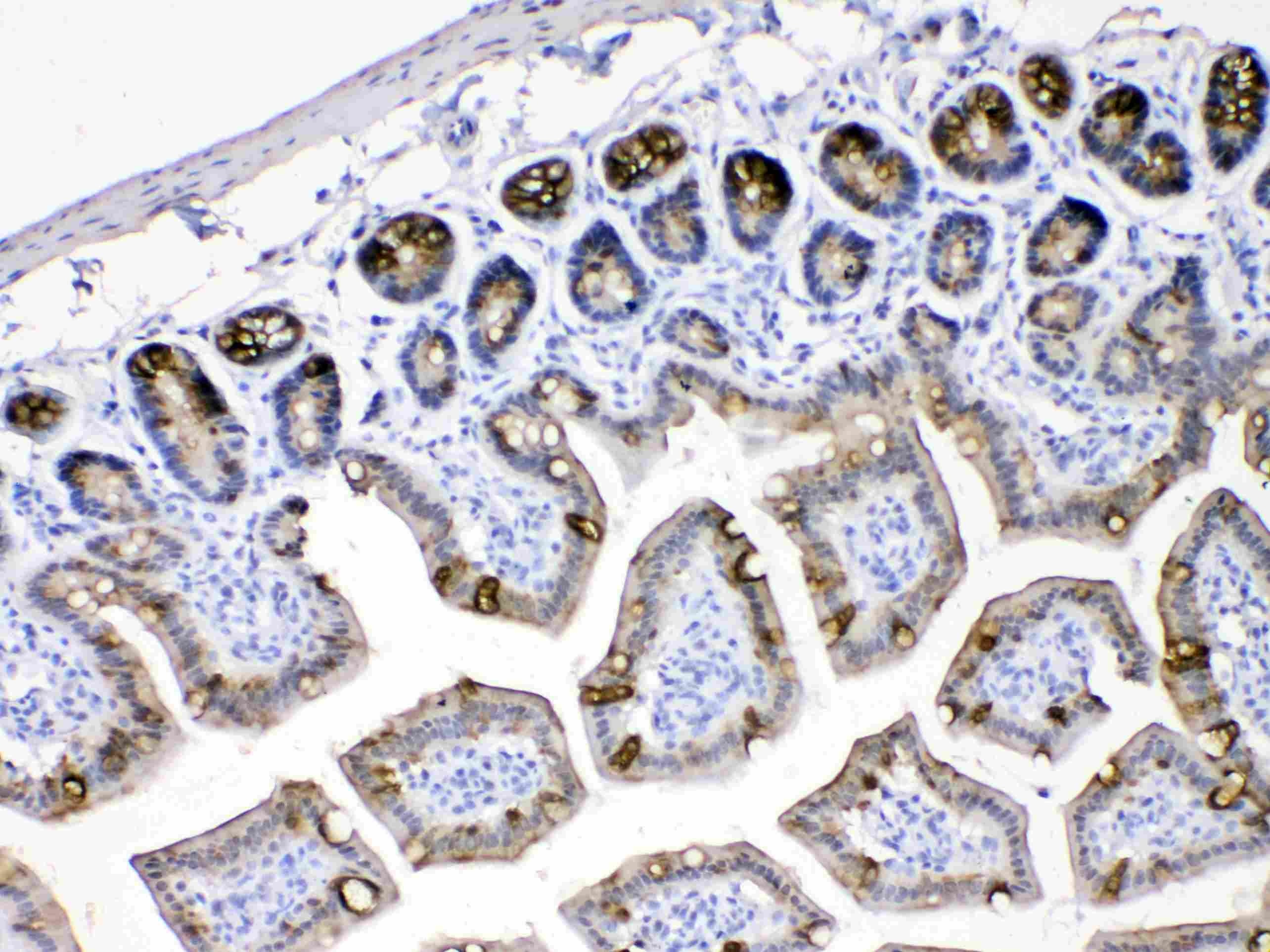
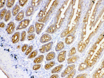
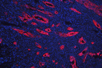
ARG40920 anti-Cytokeratin 18 antibody IHC-P image
Immunohistochemistry: Paraffin-embedded Mouse intestine tissue stained with ARG40920 anti-Cytokeratin 18 antibody at 1 µg/ml dilution.
-
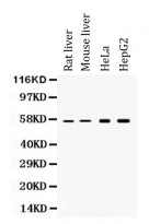
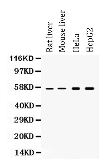
ARG40920 anti-Cytokeratin 18 antibody WB image
Western blot: Rat liver, Mouse liver, HeLa and HepG2 whole cell lysates stained with ARG40920 anti-Cytokeratin 18 antibody at 0.5 µg/ml dilution.
-
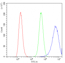
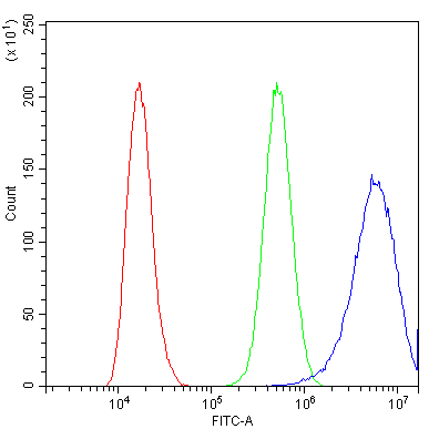
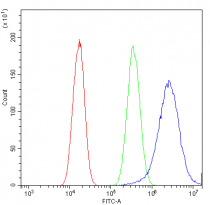
ARG40920 anti-Cytokeratin 18 antibody FACS image
Flow Cytometry: A431 cells were blocked with 10% normal goat serum and then stained with ARG40920 anti-Cytokeratin 18 antibody (blue) at 1 µg/10^6 cells for 30 min at 20°C, followed by incubation with DyLight®488 labelled secondary antibody. Isotype control antibody (green) was Rabbit IgG (1 µg/10^6 cells) used under the same conditions. Unlabelled sample (red) was also used as a control.
-
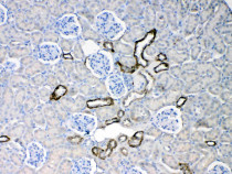
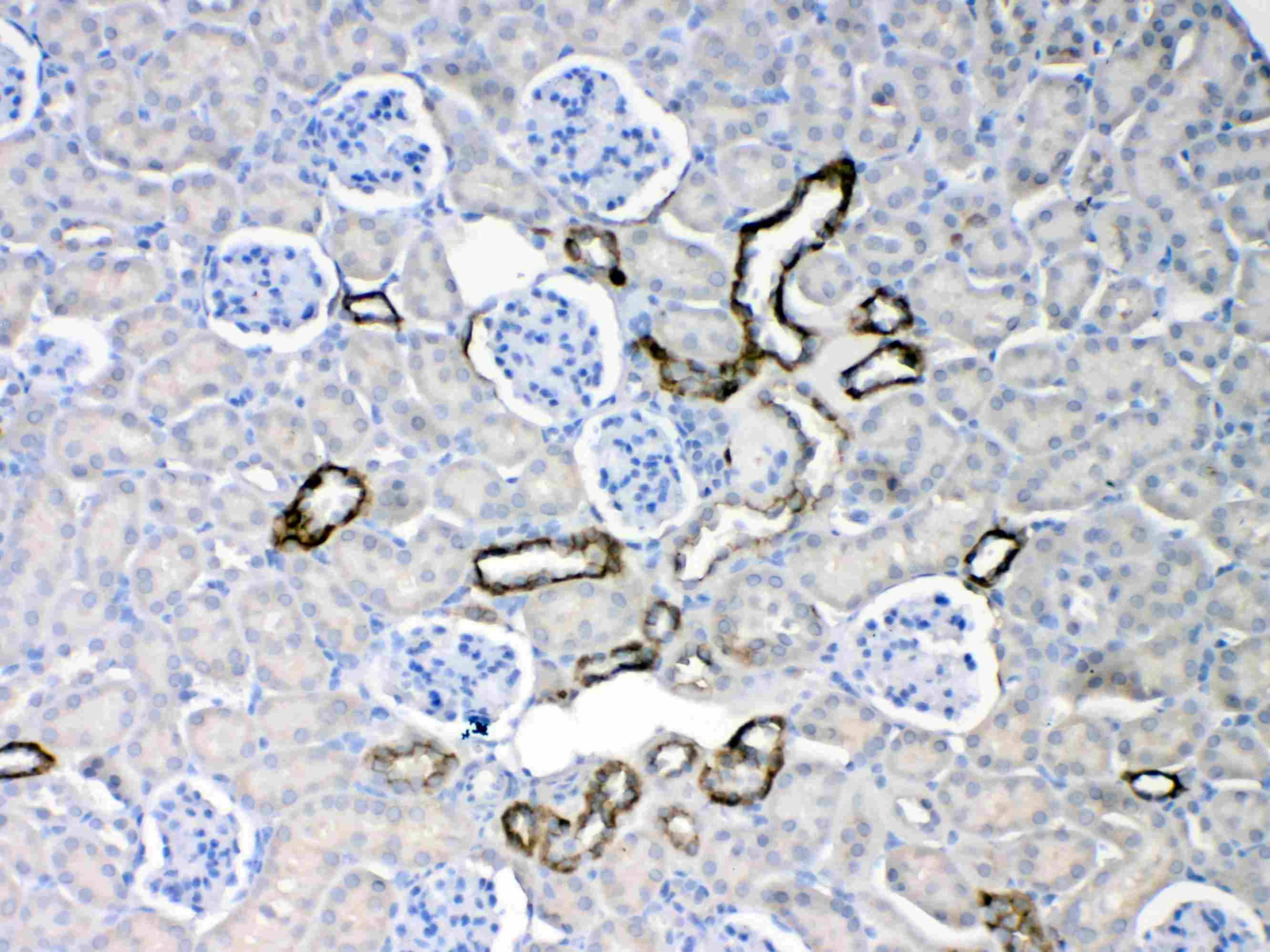
ARG40920 anti-Cytokeratin 18 antibody IHC-P image
Immunohistochemistry: Paraffin-embedded Mouse kidney tissue stained with ARG40920 anti-Cytokeratin 18 antibody at 1 µg/ml dilution.
-
ARG40920 anti-Cytokeratin 18 antibody IHC-P image
Immunohistochemistry: Paraffin-embedded Rat intestine tissue stained with ARG40920 anti-Cytokeratin 18 antibody at 1 µg/ml dilution.
-
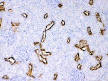
ARG40920 anti-Cytokeratin 18 antibody IHC-P image
Immunohistochemistry: Paraffin-embedded Rat kidney tissue stained with ARG40920 anti-Cytokeratin 18 antibody at 1 µg/ml dilution.
-
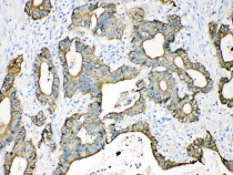
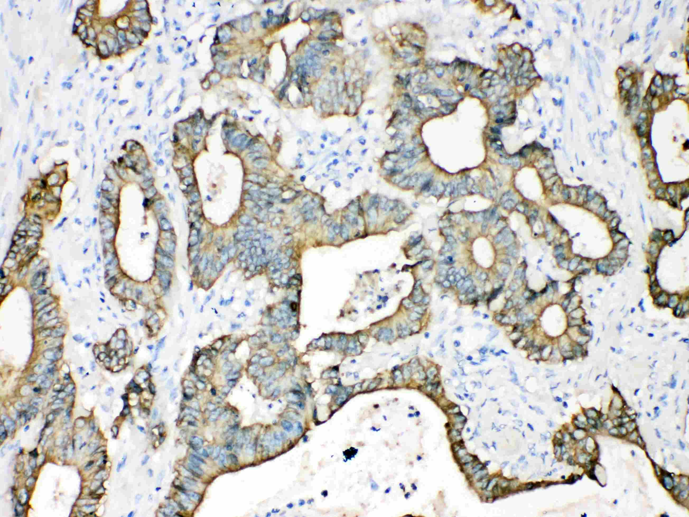
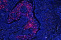
ARG40920 anti-Cytokeratin 18 antibody IHC-P image
Immunohistochemistry: Paraffin-embedded Human intetsinal cancer tissue stained with ARG40920 anti-Cytokeratin 18 antibody at 1 µg/ml dilution.
-
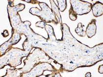
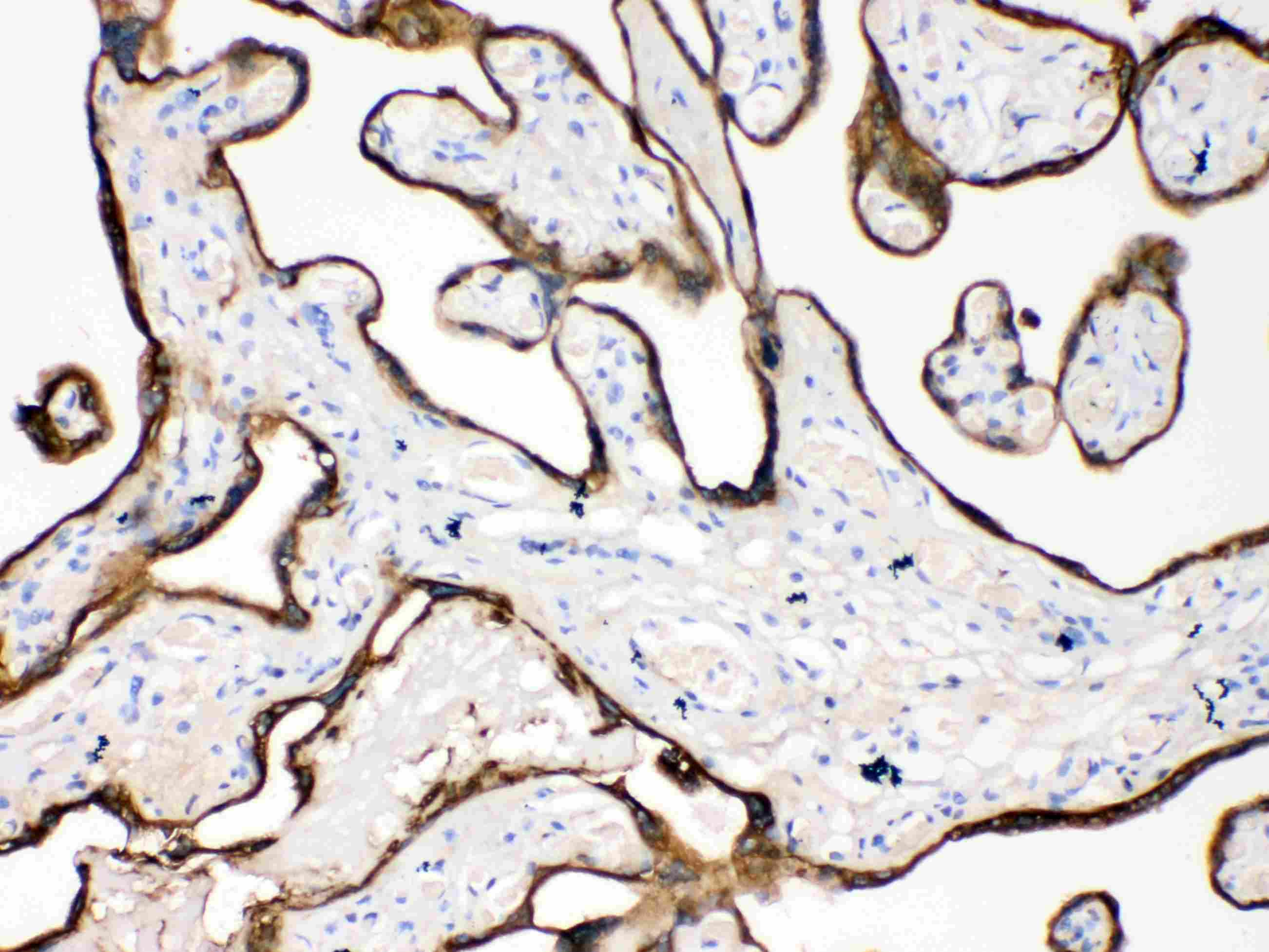
ARG40920 anti-Cytokeratin 18 antibody IHC-P image
Immunohistochemistry: Paraffin-embedded Human placenta tissue stained with ARG40920 anti-Cytokeratin 18 antibody at 1 µg/ml dilution.
-
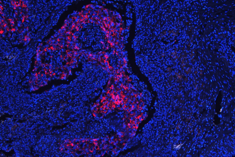
ARG40920 anti-Cytokeratin 18 antibody IHC-P image
Immunohistochemistry: Paraffin-embedded Human intestinal cancer tissue. Antigen Retrieval: Heat mediation was performed in Citrate buffer (pH 6.0, epitope retrieval solution) for 20 min. The tissue section was blocked with 10% goat serum. The tissue section was then stained with ARG40920 anti-Cytokeratin 18 antibody at 1 µg/ml, overnight at 4°C.
-
ARG40920 anti-Cytokeratin 18 antibody IHC-P image
Immunohistochemistry: Paraffin-embedded Human lung cancer tissue. Antigen Retrieval: Heat mediation was performed in Citrate buffer (pH 6.0, epitope retrieval solution) for 20 min. The tissue section was blocked with 10% goat serum. The tissue section was then stained with ARG40920 anti-Cytokeratin 18 antibody at 1 µg/ml, overnight at 4°C.
-
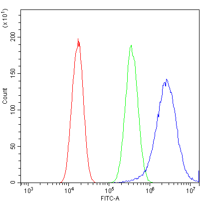
ARG40920 anti-Cytokeratin 18 antibody FACS image
Flow Cytometry: A549 cells were blocked with 10% normal goat serum and then stained with ARG40920 anti-Cytokeratin 18 antibody (blue) at 1 µg/10^6 cells for 30 min at 20°C, followed by incubation with DyLight®488 labelled secondary antibody. Isotype control antibody (green) was Rabbit IgG (1 µg/10^6 cells) used under the same conditions. Unlabelled sample (red) was also used as a control.