ARG66490
anti-Cytokeratin 13 antibody
anti-Cytokeratin 13 antibody for IHC-Formalin-fixed paraffin-embedded sections,Western blot and Human
Overview
| Product Description | Mouse Monoclonal antibody recognizes Cytokeratin 13 |
|---|---|
| Tested Reactivity | Hu |
| Tested Application | IHC-P, WB |
| Host | Mouse |
| Clonality | Monoclonal |
| Isotype | IgG1, kappa |
| Target Name | Cytokeratin 13 |
| Antigen Species | Human |
| Immunogen | Synthetic peptide derived from Human Cytokeratin 13. |
| Conjugation | Un-conjugated |
| Alternate Names | K13; Keratin, type I cytoskeletal 13; CK-13; Cytokeratin-13; WSN2; CK13; Keratin-13 |
Application Instructions
| Application Suggestion |
|
||||||
|---|---|---|---|---|---|---|---|
| Application Note | IHC-P: Antigen Retrieval: Citric acid buffer (pH 6.0) was used. * The dilutions indicate recommended starting dilutions and the optimal dilutions or concentrations should be determined by the scientist. |
Properties
| Form | Liquid |
|---|---|
| Purification | Affinity purification with immunogen. |
| Buffer | PBS, 0.02% Sodium azide, 50% Glycerol and 0.5% BSA. |
| Preservative | 0.02% Sodium azide |
| Stabilizer | 50% Glycerol and 0.5% BSA |
| Concentration | 1 mg/ml |
| Storage Instruction | For continuous use, store undiluted antibody at 2-8°C for up to a week. For long-term storage, aliquot and store at -20°C. Storage in frost free freezers is not recommended. Avoid repeated freeze/thaw cycles. Suggest spin the vial prior to opening. The antibody solution should be gently mixed before use. |
| Note | For laboratory research only, not for drug, diagnostic or other use. |
Bioinformation
| Database Links | |
|---|---|
| Gene Symbol | KRT13 |
| Gene Full Name | keratin 13, type I |
| Background | The protein encoded by this gene is a member of the keratin gene family. The keratins are intermediate filament proteins responsible for the structural integrity of epithelial cells and are subdivided into cytokeratins and hair keratins. Most of the type I cytokeratins consist of acidic proteins which are arranged in pairs of heterotypic keratin chains. This type I cytokeratin is paired with keratin 4 and expressed in the suprabasal layers of non-cornified stratified epithelia. Mutations in this gene and keratin 4 have been associated with the autosomal dominant disorder White Sponge Nevus. The type I cytokeratins are clustered in a region of chromosome 17q21.2. Alternative splicing of this gene results in multiple transcript variants; however, not all variants have been described. [provided by RefSeq, Jul 2008] |
| Calculated MW | 50 kDa |
| PTM | O-glycosylated; glycans consist of single N-acetylglucosamine residues. [UniProt] |
Images (4) Click the Picture to Zoom In
-
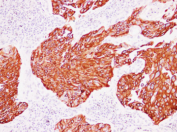
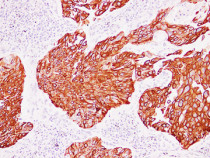
ARG66490 anti-Cytokeratin 13 antibody IHC-P image
Immunohistochemistry: Paraffin-embedded Human transitional cell carcinoma stained with ARG66490 anti-Cytokeratin 13 antibody at 1:200 (4°C, overnight). Antigen Retrieval: Citric acid buffer (pH 6.0) was used.
-
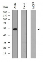
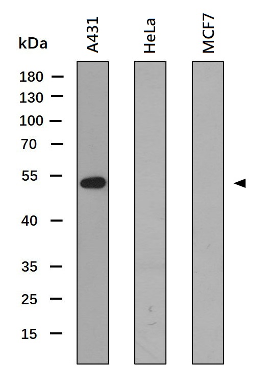
ARG66490 anti-Cytokeratin 13 antibody WB image
Western blot: 30 µg of A431, HeLa (negative control) and MCF7 (negative control) whole cell lysates stained with ARG66490 anti-Cytokeratin 13 antibody at 1:1000 dilution.
-
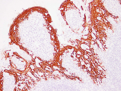
ARG66490 anti-Cytokeratin 13 antibody IHC-P image
Immunohistochemistry: Paraffin-embedded Human squamous cell lung carcinoma stained with ARG66490 anti-Cytokeratin 13 antibody at 1:200 (4°C, overnight). Antigen Retrieval: Citric acid buffer (pH 6.0) was used.
-

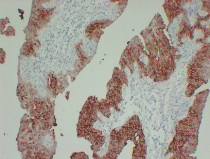
ARG66490 anti-Cytokeratin 13 antibody IHC-P image
Immunohistochemistry: Paraffin-embedded Human tonsil stained with ARG66490 anti-Cytokeratin 13 antibody at 1:200 (4°C, overnight). Antigen Retrieval: Citric acid buffer (pH 6.0) was used.