ARG20006
anti-Cytochrome C antibody
anti-Cytochrome C antibody for Immunoprecipitation,Western blot and Human,Mouse,Rat,Bovine,Chicken,Dog
Cancer antibody; Cell Biology and Cellular Response antibody; Cell Death antibody; Metabolism antibody; Signaling Transduction antibody; Mitochondria/Caspase Dependant Apoptosis Marker antibody; Mitochondrial Marker antibody; Cytochrome-C fractionation Study antibody

Overview
| Product Description | Rabbit Polyclonal antibody recognizes Cytochrome C |
|---|---|
| Tested Reactivity | Hu, Ms, Rat, Bov, Chk, Dog |
| Tested Application | IP, WB |
| Specificity | The cytochrome c antibody detects the 12.6 kDa cytochrome c from human, mouse, and rat samples. |
| Host | Rabbit |
| Clonality | Polyclonal |
| Isotype | IgG |
| Target Name | Cytochrome C |
| Antigen Species | Rat |
| Immunogen | Synthetic peptide corresponding to residues surrounding amino acid 70 of rat cytochrome C |
| Conjugation | Un-conjugated |
| Alternate Names | CYCTA |
Application Instructions
| Application Suggestion |
|
||||||
|---|---|---|---|---|---|---|---|
| Application Note | * The dilutions indicate recommended starting dilutions and the optimal dilutions or concentrations should be determined by the scientist. | ||||||
| Positive Control | Jurkat cell lysate, NIH3T3 cell lysate, and rat kidney tissue lysate |
Properties
| Form | Liquid |
|---|---|
| Purification | Affinity Purified Antibody |
| Buffer | PBS (pH 7.2), 30% Glycerol, 0.5% BSA and 0.01% Thimerosal |
| Preservative | 0.01% Thimerosal |
| Stabilizer | 30% Glycerol, 0.5% BSA |
| Concentration | 0.2 mg/ml |
| Storage Instruction | For continuous use, store undiluted antibody at 2-8°C for up to a week. For long-term storage, aliquot and store at -20°C. Storage in frost free freezers is not recommended. Avoid repeated freeze/thaw cycles. Suggest spin the vial prior to opening. The antibody solution should be gently mixed before use. |
| Note | For laboratory research only, not for drug, diagnostic or other use. |
Bioinformation
| Gene Symbol | Cyct |
|---|---|
| Gene Full Name | cytochrome c, testis |
| Background | Cytochrome c (m.w. 12,500) is an electron transport protein from mitochondria. It is released from mitochondria to cytoplasm during the early stages of apoptosis, prior to caspase activation, DNA fragmentation, and loss of membrane potential. The cytoplasmic cytochrome c is associated with Apaf-1 and caspase-9 to activate caspase-3 and other caspases |
| Function | Electron carrier protein. The oxidized form of the cytochrome c heme group can accept an electron from the heme group of the cytochrome c1 subunit of cytochrome reductase. Cytochrome c then transfers this electron to the cytochrome oxidase complex, the final protein carrier in the mitochondrial electron-transport chain. Plays a role in apoptosis. Suppression of the anti-apoptotic members or activation of the pro-apoptotic members of the Bcl-2 family leads to altered mitochondrial membrane permeability resulting in release of cytochrome c into the cytosol. Binding of cytochrome c to Apaf-1 triggers the activation of caspase-9, which then accelerates apoptosis by activating other caspases (By similarity). [UniProt] |
| Highlight | Related Antibody Duos and Panels: ARG30106 Mitochondria/Caspase Dependant Apoptosis Marker Antibody Duo (Caspase9, Cytochrome c) ARG30110 Mitochondria/Caspase dependant Apoptosis Antibody Panel (Caspase3, Caspase9, Cytochrome c, PARP) (WB) Related products: Cytochrome C antibodies; Cytochrome C Duos / Panels; Anti-Rabbit IgG secondary antibodies; Related poster download: The Structure & Functions of Mitochondria.pdf |
| Research Area | Cancer antibody; Cell Biology and Cellular Response antibody; Cell Death antibody; Metabolism antibody; Signaling Transduction antibody; Mitochondria/Caspase Dependant Apoptosis Marker antibody; Mitochondrial Marker antibody; Cytochrome-C fractionation Study antibody |
| Calculated MW | 12 kDa |
Images (1) Click the Picture to Zoom In
-
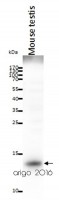
ARG20006 anti-Cytochrome C antibody WB image
Western blot: 20 µg of Mouse testis lysate stained with ARG20006 anti-Cytochrome C antibody at 2 µg/ml dilution.

Customer's Feedback
Good

Review for anti-Cytochrome C antibody
Application:WB
Sample:Mouse testis
Sample Loading Amount:20 µg
Primary Antibody Dilution Factor:2 µg/ml
Primary Antibody Incubation Time:overnight
Primary Antibody Incubation Temperature:4 ºC

Specific References
Single-Cell Analysis of Signaling Proteins Provides Insights into Proapoptotic Properties of Anticancer Drugs.
scPISA / Human


Probing apoptosis signaling proteins in single living cells for precision efficacy evaluation of anti-cancer drugs.
/ Human


