ARG40981
anti-Cyclophilin A antibody
anti-Cyclophilin A antibody for Flow cytometry,ICC/IF,Western blot and Human,Mouse,Rat
Overview
| Product Description | Rabbit Polyclonal antibody recognizes Cyclophilin A |
|---|---|
| Tested Reactivity | Hu, Ms, Rat |
| Tested Application | FACS, ICC/IF, WB |
| Host | Rabbit |
| Clonality | Polyclonal |
| Isotype | IgG |
| Target Name | Cyclophilin A |
| Antigen Species | Human |
| Immunogen | Recombinant protein corresponding to T116-E165 of Human Cyclophilin A. |
| Conjugation | Un-conjugated |
| Alternate Names | CYPA; Cyclosporin A-binding protein; PPIase A; CYPH; Cyclophilin A; HEL-S-69p; EC 5.2.1.8; Rotamase A; Peptidyl-prolyl cis-trans isomerase A |
Application Instructions
| Application Suggestion |
|
||||||||
|---|---|---|---|---|---|---|---|---|---|
| Application Note | * The dilutions indicate recommended starting dilutions and the optimal dilutions or concentrations should be determined by the scientist. |
Properties
| Form | Liquid |
|---|---|
| Purification | Affinity purification with immunogen. |
| Buffer | 0.2% Na2HPO4, 0.9% NaCl, 0.05% Sodium azide and 4% Trehalose. |
| Preservative | 0.05% Sodium azide |
| Stabilizer | 4% Trehalose |
| Concentration | 0.5 mg/ml |
| Storage Instruction | For continuous use, store undiluted antibody at 2-8°C for up to a week. For long-term storage, aliquot and store at -20°C or below. Storage in frost free freezers is not recommended. Avoid repeated freeze/thaw cycles. Suggest spin the vial prior to opening. The antibody solution should be gently mixed before use. |
| Note | For laboratory research only, not for drug, diagnostic or other use. |
Bioinformation
| Database Links | |
|---|---|
| Gene Symbol | PPIA |
| Gene Full Name | peptidylprolyl isomerase A (cyclophilin A) |
| Background | This gene encodes a member of the peptidyl-prolyl cis-trans isomerase (PPIase) family. PPIases catalyze the cis-trans isomerization of proline imidic peptide bonds in oligopeptides and accelerate the folding of proteins. The encoded protein is a cyclosporin binding-protein and may play a role in cyclosporin A-mediated immunosuppression. The protein can also interact with several HIV proteins, including p55 gag, Vpr, and capsid protein, and has been shown to be necessary for the formation of infectious HIV virions. Multiple pseudogenes that map to different chromosomes have been reported. [provided by RefSeq, Jul 2008] |
| Function | PPIases accelerate the folding of proteins. It catalyzes the cis-trans isomerization of proline imidic peptide bonds in oligopeptides. [UniProt] |
| Cellular Localization | Cytoplasm. Secreted. Note=Secretion occurs in response to oxidative stress in vascular smooth muscle through a vesicular secretory pathway that involves actin remodeling and myosin II activation, and mediates ERK1/2 activation. [UniProt] |
| Calculated MW | 18 kDa |
| PTM | Acetylation at Lys-125 markedly inhibits catalysis of cis to trans isomerization and stabilizes cis rather than trans forms of the HIV-1 capsid. PPIA acetylation also antagonizes the immunosuppressive effects of cyclosporine by inhibiting the sequential steps of cyclosporine binding and calcineurin inhibition. [UniProt] |
Images (5) Click the Picture to Zoom In
-

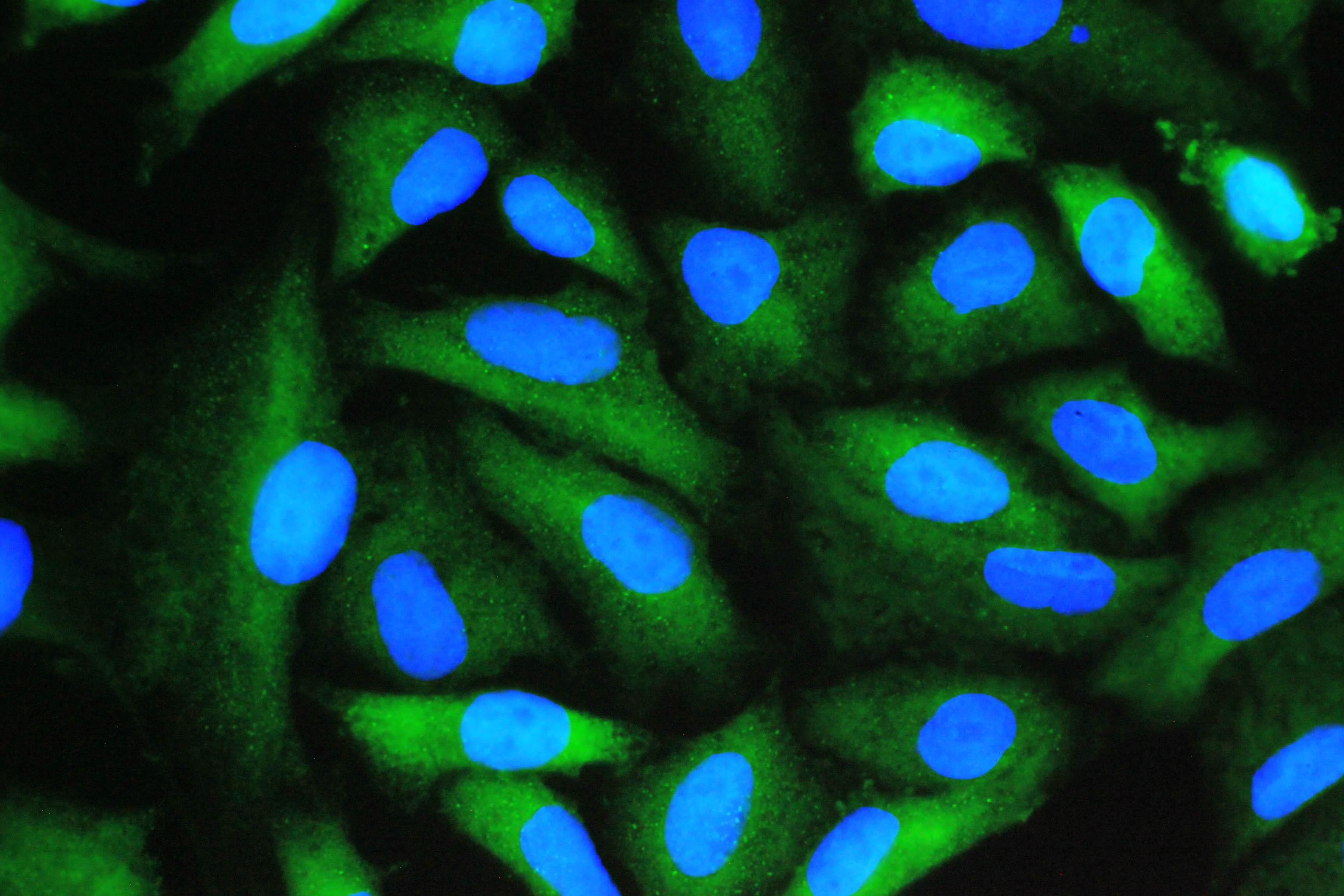
ARG40981 anti-Cyclophilin A antibody ICC/IF image
Immunofluorescence: A549 cells stained with ARG40981 anti-Cyclophilin A antibody (green) at 2 µg/ml, overnight at 4°C. DAPI (blue) for nuclear staining.
-

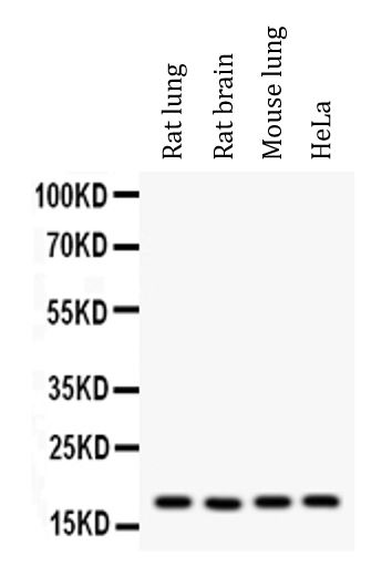
ARG40981 anti-Cyclophilin A antibody WB image
Western blot: 50 µg of samples under reducing conditions. Rat lung, Rat brain, Mouse lung and HeLa whole cell lysates stained with ARG40981 anti-Cyclophilin A antibody at 0.5 µg/ml, overnight at 4°C.
-
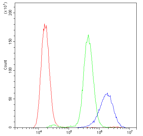
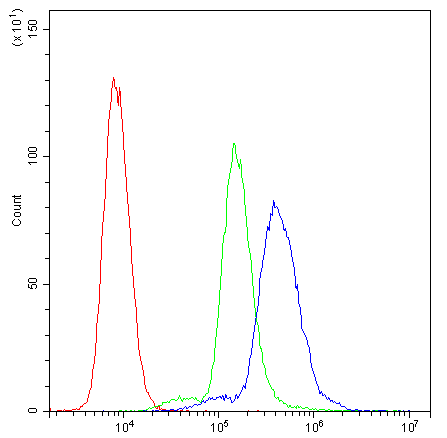
ARG40981 anti-Cyclophilin A antibody FACS image
Flow Cytometry: THP-1 cells were blocked with 10% normal goat serum and then stained with ARG40981 anti-Cyclophilin A antibody (blue) at 1 µg/10^6 cells for 30 min at 20°C, followed by incubation with DyLight®488 labelled secondary antibody. Isotype control antibody (green) was Rabbit IgG (1 µg/10^6 cells) used under the same conditions. Unlabelled sample (red) was also used as a control.
-
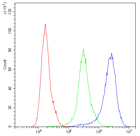
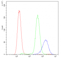
ARG40981 anti-Cyclophilin A antibody FACS image
Flow Cytometry: U937 cells were blocked with 10% normal goat serum and then stained with ARG40981 anti-Cyclophilin A antibody (blue) at 1 µg/10^6 cells for 30 min at 20°C, followed by incubation with DyLight®488 labelled secondary antibody. Isotype control antibody (green) was Rabbit IgG (1 µg/10^6 cells) used under the same conditions. Unlabelled sample (red) was also used as a control.
-
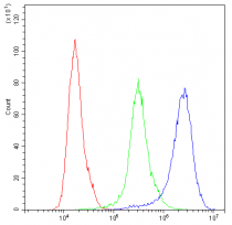
ARG40981 anti-Cyclophilin A antibody FACS image
Flow Cytometry: K562 cells were blocked with 10% normal goat serum and then stained with ARG40981 anti-Cyclophilin A antibody (blue) at 1 µg/10^6 cells for 30 min at 20°C, followed by incubation with DyLight®488 labelled secondary antibody. Isotype control antibody (green) was Rabbit IgG (1 µg/10^6 cells) used under the same conditions. Unlabelled sample (red) was also used as a control.