ARG58314
anti-Cyclin T1 antibody
anti-Cyclin T1 antibody for Flow cytometry,ICC/IF,IHC-Formalin-fixed paraffin-embedded sections,Western blot and Human,Mouse,Rat
Overview
| Product Description | Rabbit Polyclonal antibody recognizes Cyclin T1 |
|---|---|
| Tested Reactivity | Hu, Ms, Rat |
| Predict Reactivity | Bov |
| Tested Application | FACS, ICC/IF, IHC-P, WB |
| Host | Rabbit |
| Clonality | Polyclonal |
| Isotype | IgG |
| Target Name | Cyclin T1 |
| Antigen Species | Human |
| Immunogen | Synthetic peptide corresponding to a sequence in the middle region of Human Cyclin T1 (375-410aa QKQNSKSVPSAKVSLKEYRAKHAEELAAQKRQLENM), different from the related Mouse sequence by one amino acid. |
| Conjugation | Un-conjugated |
| Alternate Names | CCNT; Cyclin-T; HIVE1; CYCT1; CycT1; Cyclin-T1 |
Application Instructions
| Application Suggestion |
|
||||||||||
|---|---|---|---|---|---|---|---|---|---|---|---|
| Application Note | IHC-P: Antigen Retrieval: By heat mediation. * The dilutions indicate recommended starting dilutions and the optimal dilutions or concentrations should be determined by the scientist. |
Properties
| Form | Liquid |
|---|---|
| Purification | Affinity purification with immunogen. |
| Buffer | 0.9% NaCl, 0.2% Na2HPO4, 0.05% Sodium azide and 5% BSA. |
| Preservative | 0.05% Sodium azide |
| Stabilizer | 5% BSA |
| Concentration | 0.5 mg/ml |
| Storage Instruction | For continuous use, store undiluted antibody at 2-8°C for up to a week. For long-term storage, aliquot and store at -20°C or below. Storage in frost free freezers is not recommended. Avoid repeated freeze/thaw cycles. Suggest spin the vial prior to opening. The antibody solution should be gently mixed before use. |
| Note | For laboratory research only, not for drug, diagnostic or other use. |
Bioinformation
| Database Links | |
|---|---|
| Gene Symbol | CCNT1 |
| Gene Full Name | cyclin T1 |
| Background | This gene encodes a member of the highly conserved cyclin C subfamily. The encoded protein tightly associates with cyclin-dependent kinase 9, and is a major subunit of positive transcription elongation factor b (p-TEFb). In humans, there are multiple forms of positive transcription elongation factor b, which may include one of several different cyclins along with cyclin-dependent kinase 9. The complex containing the encoded cyclin and cyclin-dependent kinase 9 acts as a cofactor of human immunodeficiency virus type 1 (HIV-1) Tat protein, and is both necessary and sufficient for full activation of viral transcription. This cyclin and its kinase partner are also involved in triggering transcript elongation through phosphorylation of the carboxy-terminal domain of the largest RNA polymerase II subunit. Overexpression of this gene is implicated in tumor growth. Alternative splicing results in multiple transcript variants. [provided by RefSeq, Apr 2013] |
| Function | Regulatory subunit of the cyclin-dependent kinase pair (CDK9/cyclin-T1) complex, also called positive transcription elongation factor B (P-TEFb), which is proposed to facilitate the transition from abortive to productive elongation by phosphorylating the CTD (carboxy-terminal domain) of the large subunit of RNA polymerase II (RNA Pol II). In case of HIV or SIV infections, binds to the transactivation domain of the viral nuclear transcriptional activator, Tat, thereby increasing Tat's affinity for the transactivating response RNA element (TAR RNA). Serves as an essential cofactor for Tat, by promoting RNA Pol II activation, allowing transcription of viral genes. [UniProt] |
| Cellular Localization | Nucleus. [UniProt] |
| Calculated MW | 81 kDa |
Images (7) Click the Picture to Zoom In
-
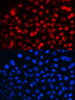
ARG58314 anti-Cyclin T1 antibody ICC/IF image
Immunofluorescence: A431 cells were blocked with 10% goat serum and then stained with ARG58314 anti-Cyclin T1 antibody (red) at 2 µg/ml dilution, overnight at 4°C. DAPI (blue) for nuclear staining.
-
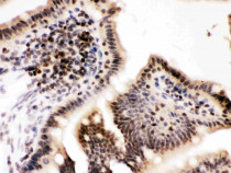
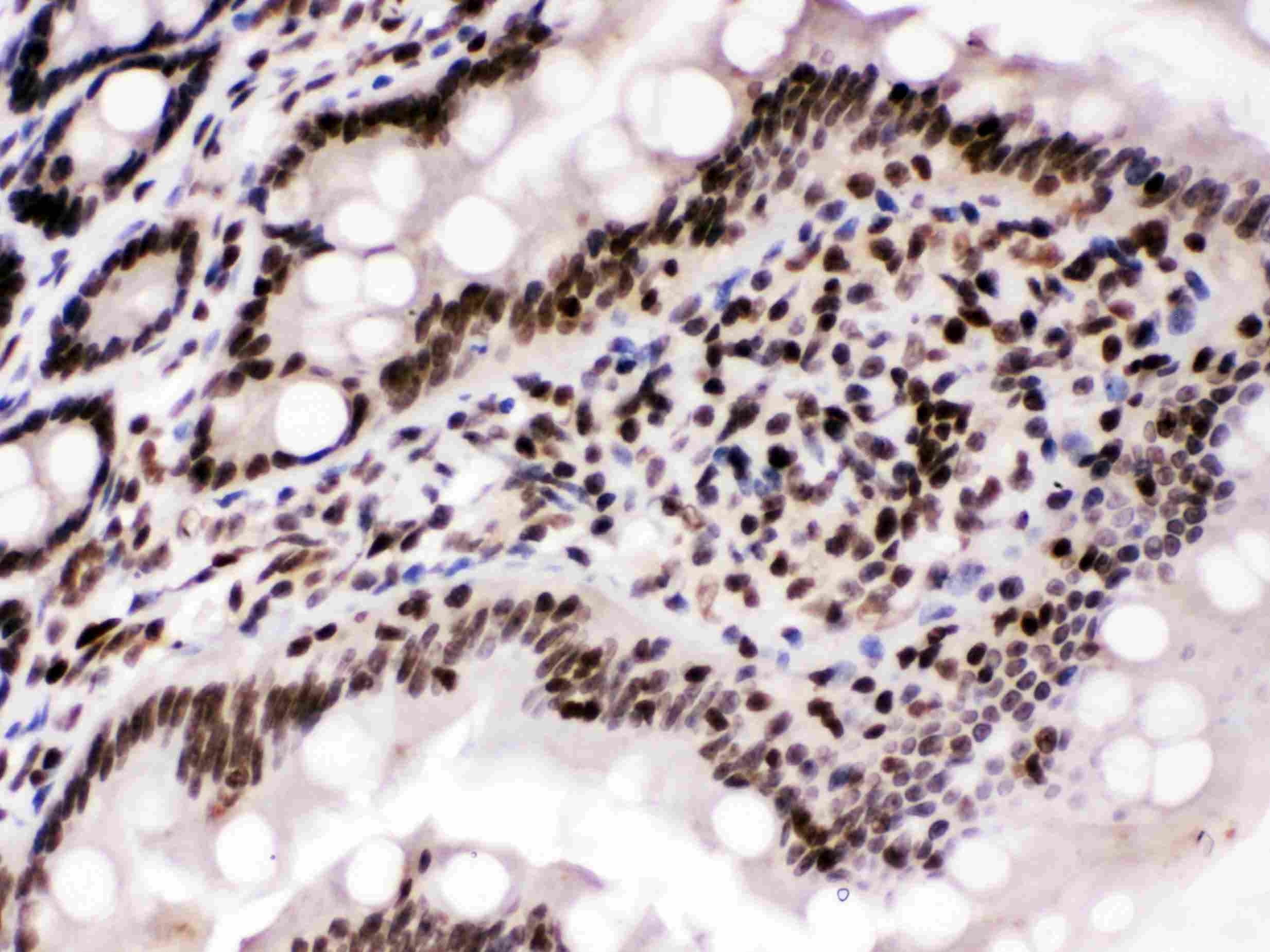
ARG58314 anti-Cyclin T1 antibody IHC-P image
Immunohistochemistry: Paraffin-embedded Mouse intestine stained with ARG58314 anti-Cyclin T1 antibody at 1 µg/ml dilution.
-

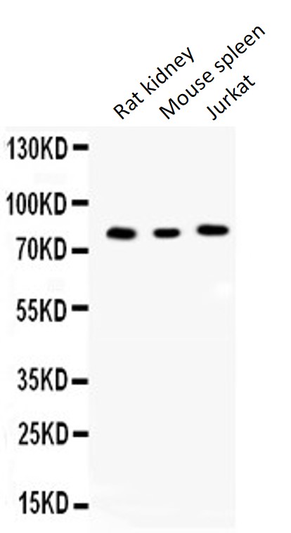
ARG58314 anti-Cyclin T1 antibody WB image
Western blot: Rat kidney extract, Mouse spleen extract and Jurkat whole cell lysates stained with ARG58314 anti-Cyclin T1 antibody at 0.5 µg/ml dilution.
-

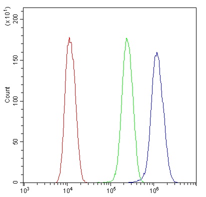
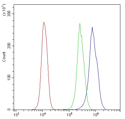
ARG58314 anti-Cyclin T1 antibody FACS image
Flow Cytometry: U2OS cells were blocked with 10% normal goat serum and then stained with ARG58314 anti-Cyclin T1 antibody (blue) at 1 µg/10^6 cells for 30 min at 20°C, followed by incubation with DyLight®488 labelled secondary antibody. Isotype control antibody (green) was rabbit IgG (1 µg/10^6 cells) used under the same conditions. Unlabelled sample (red) was also used as a control.
-

ARG58314 anti-Cyclin T1 antibody IHC-P image
Immunohistochemistry: Paraffin-embedded Rat intestine stained with ARG58314 anti-Cyclin T1 antibody at 1 µg/ml dilution.
-
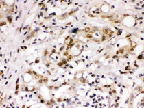
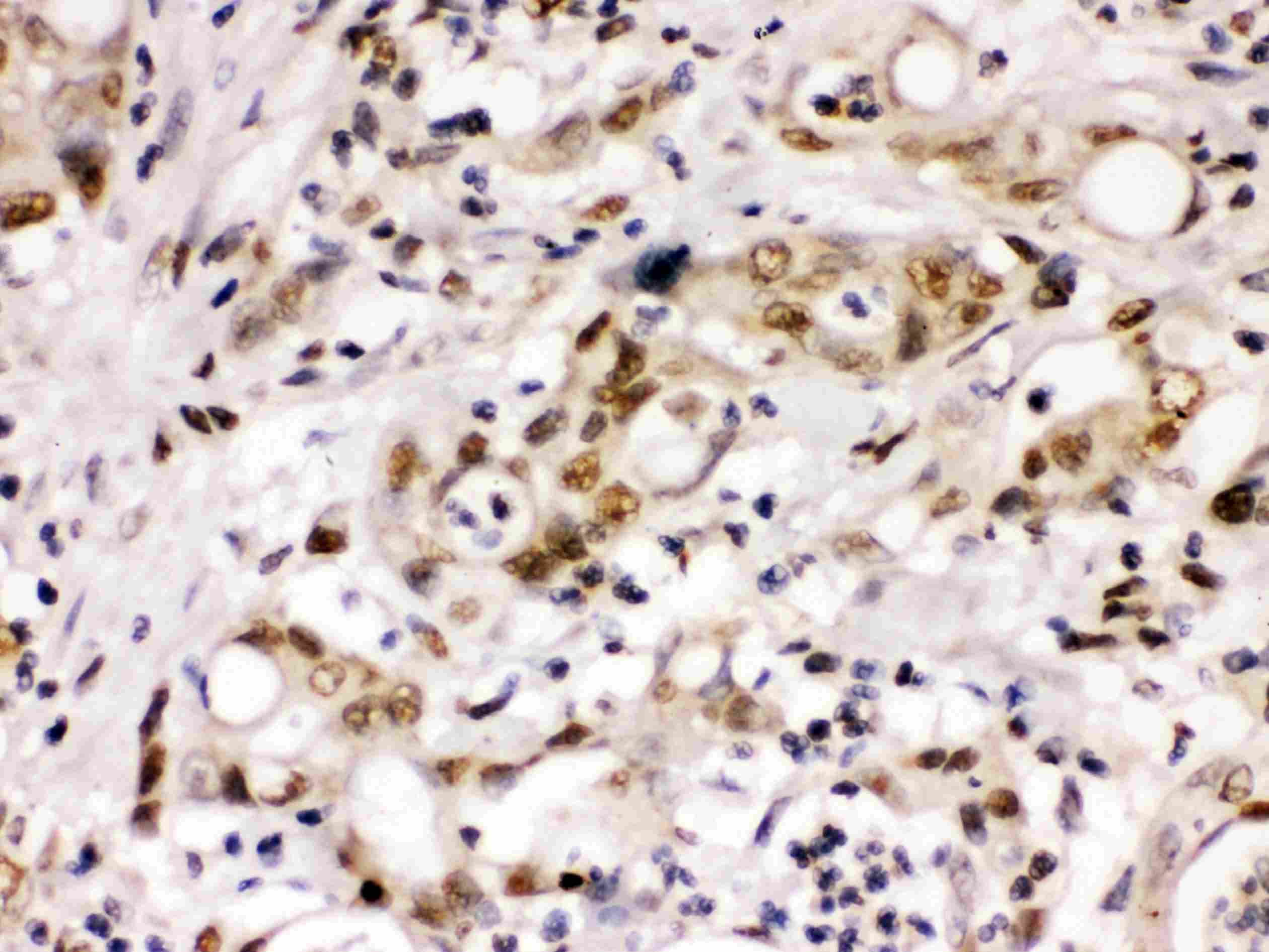
ARG58314 anti-Cyclin T1 antibody IHC-P image
Immunohistochemistry: Paraffin-embedded Human intestinal cancer stained with ARG58314 anti-Cyclin T1 antibody at 1 µg/ml dilution.
-
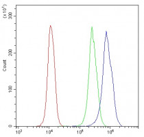
ARG58314 anti-Cyclin T1 antibody FACS image
Flow Cytometry: U937 cells were blocked with 10% normal goat serum and then stained with ARG58314 anti-Cyclin T1 antibody (blue) at 1 µg/10^6 cells for 30 min at 20°C, followed by incubation with DyLight®488 labelled secondary antibody. Isotype control antibody (green) was rabbit IgG (1 µg/10^6 cells) used under the same conditions. Unlabelled sample (red) was also used as a control.