ARG55933
anti-Cyclin D1 antibody [DCS-6]
anti-Cyclin D1 antibody [DCS-6] for Flow cytometry,ICC/IF,IHC-Formalin-fixed paraffin-embedded sections,Western blot and Human,Mouse,Rat
Overview
| Product Description | Mouse Monoclonal antibody [DCS-6] recognizes Cyclin D1 |
|---|---|
| Tested Reactivity | Hu, Ms, Rat |
| Tested Application | FACS, ICC/IF, IHC-P, WB |
| Host | Mouse |
| Clonality | Monoclonal |
| Clone | DCS-6 |
| Isotype | IgG2a, kappa |
| Target Name | Cyclin D1 |
| Antigen Species | Human |
| Immunogen | Human recombinant full length Cyclin D1 protein. |
| Conjugation | Un-conjugated |
| Alternate Names | B-cell lymphoma 1 protein; PRAD1; U21B31; D11S287E; BCL-1; G1/S-specific cyclin-D1; BCL-1 oncogene; BCL1; PRAD1 oncogene |
Application Instructions
| Application Suggestion |
|
||||||||||
|---|---|---|---|---|---|---|---|---|---|---|---|
| Application Note | IHC-P: Antigen Retrieval: Boil tissue section in 10 mM Citrate buffer (pH 6.0) for 10-20 min, followed by cooling at RT for 20 min. * The dilutions indicate recommended starting dilutions and the optimal dilutions or concentrations should be determined by the scientist. |
Properties
| Form | Liquid |
|---|---|
| Purification | Purification with Protein G. |
| Buffer | PBS (pH 7.4), 0.05% Sodium azide and 0.1 mg/ml BSA |
| Preservative | 0.05% Sodium azide |
| Stabilizer | 0.1 mg/ml BSA |
| Concentration | 0.2 mg/ml |
| Storage Instruction | For continuous use, store undiluted antibody at 2-8°C for up to a week. For long-term storage, aliquot and store at -20°C or below. Storage in frost free freezers is not recommended. Avoid repeated freeze/thaw cycles. Suggest spin the vial prior to opening. The antibody solution should be gently mixed before use. |
| Note | For laboratory research only, not for drug, diagnostic or other use. |
Bioinformation
| Database Links | |
|---|---|
| Gene Symbol | CCND1 |
| Gene Full Name | cyclin D1 |
| Background | The protein encoded by this gene belongs to the highly conserved cyclin family, whose members are characterized by a dramatic periodicity in protein abundance throughout the cell cycle. Cyclins function as regulators of CDK kinases. Different cyclins exhibit distinct expression and degradation patterns which contribute to the temporal coordination of each mitotic event. This cyclin forms a complex with and functions as a regulatory subunit of CDK4 or CDK6, whose activity is required for cell cycle G1/S transition. This protein has been shown to interact with tumor suppressor protein Rb and the expression of this gene is regulated positively by Rb. Mutations, amplification and overexpression of this gene, which alters cell cycle progression, are observed frequently in a variety of tumors and may contribute to tumorigenesis. [provided by RefSeq, Jul 2008] |
| Function | Regulatory component of the cyclin D1-CDK4 (DC) complex that phosphorylates and inhibits members of the retinoblastoma (RB) protein family including RB1 and regulates the cell-cycle during G(1)/S transition. Phosphorylation of RB1 allows dissociation of the transcription factor E2F from the RB/E2F complex and the subsequent transcription of E2F target genes which are responsible for the progression through the G(1) phase. Hypophosphorylates RB1 in early G(1) phase. Cyclin D-CDK4 complexes are major integrators of various mitogenenic and antimitogenic signals. Also substrate for SMAD3, phosphorylating SMAD3 in a cell-cycle-dependent manner and repressing its transcriptional activity. Component of the ternary complex, cyclin D1/CDK4/CDKN1B, required for nuclear translocation and activity of the cyclin D-CDK4 complex. Exhibits transcriptional corepressor activity with INSM1 on the NEUROD1 and INS promoters in a cell cycle-independent manner. [UniProt] |
| Cellular Localization | Nuclear |
| Calculated MW | 34 kDa |
| PTM | Phosphorylation at Thr-286 by MAP kinases is required for ubiquitination and degradation following DNA damage. It probably plays an essential role for recognition by the FBXO31 component of SCF (SKP1-cullin-F-box) protein ligase complex. Ubiquitinated, primarily as 'Lys-48'-linked polyubiquitination. Ubiquitinated by a SCF (SKP1-CUL1-F-box protein) ubiquitin-protein ligase complex containing FBXO4 and CRYAB. Following DNA damage it is ubiquitinated by some SCF (SKP1-cullin-F-box) protein ligase complex containing FBXO31. SCF-type ubiquitination is dependent on Thr-286 phosphorylation (By similarity). Ubiquitinated also by UHRF2 apparently in a phosphorylation-independent manner. Ubiquitination leads to its degradation and G1 arrest. Deubiquitinated by USP2; leading to its stabilization. |
Images (4) Click the Picture to Zoom In
-
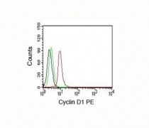
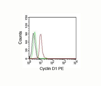
ARG55933 anti-Cyclin D1 antibody [DCS-6] FACS image
Flow Cytometry: Jurkat cells stained with PE-conjugate ARG55933 anti-Cyclin D1 antibody [DCS-6] (red); Cells alone (black); Isotype control (green).
-
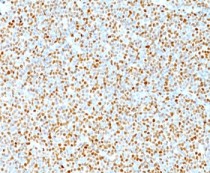
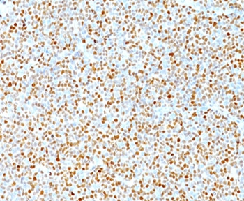
ARG55933 anti-Cyclin D1 antibody [DCS-6] IHC-P image
Immunohistochemistry: Human mantle cell lymphoma stained with ARG55933 anti-Cyclin D1 antibody [DCS-6]. Nuclear tumor cells is seen.
-
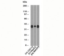
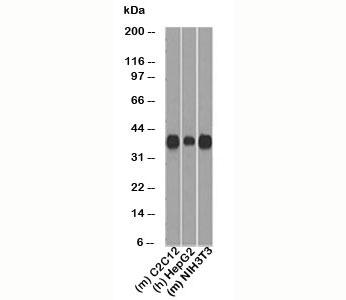
ARG55933 anti-Cyclin D1 antibody [DCS-6] WB image
Western blot: C2C12, HepG2 and NIH3T3 cell lysates stained with ARG55933 anti-Cyclin D1 antibody [DCS-6]. Predicted molecular weight: 32-36 kDa.
-
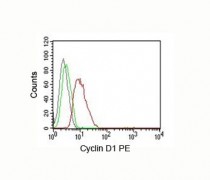
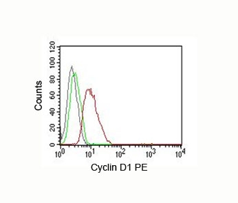
ARG55933 anti-Cyclin D1 antibody [DCS-6] FACS image
Flow Cytometry: MCF-7 cells stained with PE-conjugate ARG55933 anti-Cyclin D1 antibody [DCS-6] (red); Cells alone (black); Isotype control (green).