ARG10084
anti-Colorectal Carcinoma / CD3 antibody [BS-1]
anti-Colorectal Carcinoma / CD3 antibody [BS-1] for ELISA,Flow cytometry and Human
Controls and Markers antibody; Colorectal Carcinoma Marker antibody
Overview
| Product Description | Mouse Monoclonal antibody [BS-1] recognizes colorectal carcinoma / CD3. This is a bispecific antibody produced by fusion of two hybridoma cell lines. The human colorectal carcinoma antigen (CRC) mAb secreting cell line was transfected by mpSV2gpt. The human CD3 mAb secreting cell line (JXT3) was transfected by mpSV2neo. The somatic fusion between CRC and JXT3 cells produced quadroma CRCgpt / CD3neo, which were selected and cloned in media containing both Mycophenolic acid and Geneticin. Quadromas showing both murine IgG1 and IgG2a was subcloned for bispecific antibody CRC / CD3 production. |
|---|---|
| Tested Reactivity | Hu |
| Tested Application | ELISA, FACS |
| Host | Mouse |
| Clonality | Monoclonal |
| Clone | BS-1 |
| Isotype | IgG1 / IgG2a |
| Target Name | Colorectal Carcinoma / CD3 |
| Antigen Species | Human |
| Immunogen | The original CRC mAb (Y94) used human colorectal carcinoma as immunogen. The original CD3 mAb (JXT3) used human peripheral T lymphocytes as immunogen |
| Conjugation | Un-conjugated |
Application Instructions
| Application Note | ELISA: The bi-specific antibody has been shown to detect the presence of the tumor-associated antigen in the serum of patients with colorectal carcinoma, and reacted with mucin-like oncofetal pancarcinoma antigen, glycoprotein TAG-72. * The dilutions indicate recommended starting dilutions and the optimal dilutions or concentrations should be determined by the scientist. |
|---|
Properties
| Form | Liquid |
|---|---|
| Purification | Protein G affinity purified |
| Buffer | 0.01M PBS (pH 7.0) |
| Concentration | 1 mg/ml |
| Storage Instruction | For continuous use, store undiluted antibody at 2-8°C for up to a week. For long-term storage, aliquot and store at -20°C or below. Storage in frost free freezers is not recommended. Avoid repeated freeze/thaw cycles. Suggest spin the vial prior to opening. The antibody solution should be gently mixed before use. |
| Note | For laboratory research only, not for drug, diagnostic or other use. |
Bioinformation
| Background | Colorectal carcinoma is the cancer developed in the colon or rectum of the digestive system. In developed countries, it is the most common cancer in aging population. Genetic deposition and a less active life style contribute to the development of the cancer. Molecular pathological study showed that altered Wnt-APC-β-catenim signaling pathway, mutated p53, and deactivated TGF-β and DCC (Deleted in Colon Cancer) are involved with the pathogenesis. The cancer is currently screened with a fecal occult blood test in people over 50 years old and the malignancy is confirmed by tumor biopsy. The search for specific biomarker for non-invasive test is still ongoing. CD3 exists on the cell surface of all T-cell types. It is used for differentiating T-cells from other leukocytes such as B cells and natural killer cells. CD3 is the accessory molecule in the T cell receptor complex. In the presence of CD3 and ζ-chain, T-cell receptor binds to antigen presented by MHC and transfers signal for T-cell activation. The hybrid bi-specific antibody binds to CD3 and colorectal carcinoma related antigen at its two different Fabs. Theoretically, the bi-specific antibody brings the target cancer antigen near T-cells and could enhance T-cell mediated immunity to cancer. However, if the binding to CD3 disrupts the CD3’s accessory function, T-cell immunity suppression could be resulted. |
|---|---|
| Research Area | Controls and Markers antibody; Colorectal Carcinoma Marker antibody |
Images (1) Click the Picture to Zoom In
-
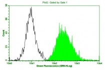
ARG10084 anti-Colorectal Carcinoma/CD3 antibody [BS-1] Flow Cytometry image
Flow Cytometry: Jurkat cells stained with anti-Colorectal Carcinoma/CD3 antibody [BS-1] (ARG10084) followed by incubation with fluorescence labelled secondary antibody.


