ARG66486
anti-Claudin 3 antibody
anti-Claudin 3 antibody for IHC-Formalin-fixed paraffin-embedded sections,Western blot and Human
Overview
| Product Description | Mouse Monoclonal antibody recognizes Claudin 3 |
|---|---|
| Tested Reactivity | Hu |
| Tested Application | IHC-P, WB |
| Host | Mouse |
| Clonality | Monoclonal |
| Isotype | IgG1, kappa |
| Target Name | Claudin 3 |
| Antigen Species | Human |
| Immunogen | Synthetic peptide derived from Human Claudin 3. |
| Conjugation | Un-conjugated |
| Alternate Names | CPE-R2; CPE-receptor 2; RVP1; HRVP1; CPE-R 2; CPETR2; hRVP1; C7orf1; Clostridium perfringens enterotoxin receptor 2; Rat ventral prostate.1 protein homolog; Claudin-3 |
Application Instructions
| Application Suggestion |
|
||||||
|---|---|---|---|---|---|---|---|
| Application Note | IHC-P: Antigen Retrieval: Citric acid buffer (pH 6.0) was used. * The dilutions indicate recommended starting dilutions and the optimal dilutions or concentrations should be determined by the scientist. |
||||||
| Observed Size | ~ 22 kDa |
Properties
| Form | Liquid |
|---|---|
| Purification | Affinity purification with immunogen. |
| Buffer | PBS, 0.02% Sodium azide, 50% Glycerol and 0.5% BSA. |
| Preservative | 0.02% Sodium azide |
| Stabilizer | 50% Glycerol and 0.5% BSA |
| Concentration | 1 mg/ml |
| Storage Instruction | For continuous use, store undiluted antibody at 2-8°C for up to a week. For long-term storage, aliquot and store at -20°C. Storage in frost free freezers is not recommended. Avoid repeated freeze/thaw cycles. Suggest spin the vial prior to opening. The antibody solution should be gently mixed before use. |
| Note | For laboratory research only, not for drug, diagnostic or other use. |
Bioinformation
| Database Links | |
|---|---|
| Gene Symbol | CLDN3 |
| Gene Full Name | claudin 3 |
| Background | Tight junctions represent one mode of cell-to-cell adhesion in epithelial or endothelial cell sheets, forming continuous seals around cells and serving as a physical barrier to prevent solutes and water from passing freely through the paracellular space. These junctions are comprised of sets of continuous networking strands in the outwardly facing cytoplasmic leaflet, with complementary grooves in the inwardly facing extracytoplasmic leaflet. The protein encoded by this intronless gene, a member of the claudin family, is an integral membrane protein and a component of tight junction strands. It is also a low-affinity receptor for Clostridium perfringens enterotoxin, and shares aa sequence similarity with a putative apoptosis-related protein found in rat. [provided by RefSeq, Jul 2008] |
| Function | Plays a major role in tight junction-specific obliteration of the intercellular space, through calcium-independent cell-adhesion activity. [UniProt] |
| Cellular Localization | Cell junction, tight junction. Cell membrane; Multi-pass membrane protein. [UniProt] |
| Calculated MW | 23 kDa |
Images (5) Click the Picture to Zoom In
-
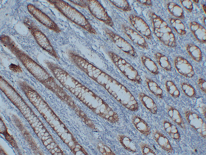
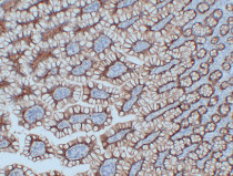
ARG66486 anti-Claudin 3 antibody IHC-P image
Immunohistochemistry: Paraffin-embedded Human colon stained with ARG66486 anti-Claudin 3 antibody at 1:200 (4°C, overnight). Antigen Retrieval: Citric acid buffer (pH 6.0) was used.
-

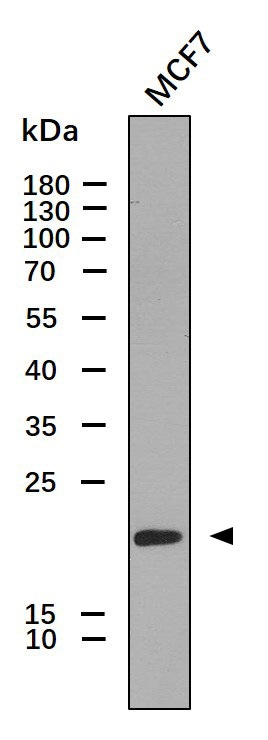
ARG66486 anti-Claudin 3 antibody WB image
Western blot: 30 µg of MCF7 whole cell lysate stained with ARG66486 anti-Claudin 3 antibody at 1:1000 dilution.
-
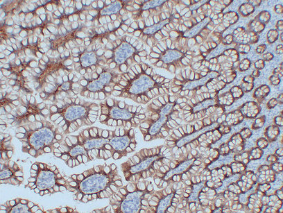
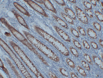
ARG66486 anti-Claudin 3 antibody IHC-P image
Immunohistochemistry: Paraffin-embedded Human intestine stained with ARG66486 anti-Claudin 3 antibody at 1:200 (4°C, overnight). Antigen Retrieval: Citric acid buffer (pH 6.0) was used.
-
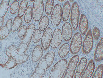
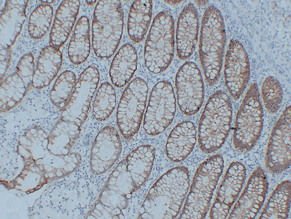
ARG66486 anti-Claudin 3 antibody IHC-P image
Immunohistochemistry: Paraffin-embedded Human rectum stained with ARG66486 anti-Claudin 3 antibody at 1:200 (4°C, overnight). Antigen Retrieval: Citric acid buffer (pH 6.0) was used.
-
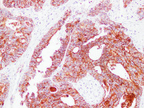
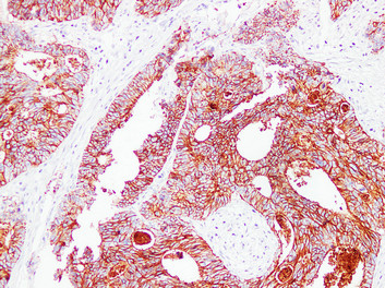
ARG66486 anti-Claudin 3 antibody IHC-P image
Immunohistochemistry: Paraffin-embedded Human colon carcinoma stained with ARG66486 anti-Claudin 3 antibody at 1:200 (4°C, overnight). Antigen Retrieval: Citric acid buffer (pH 6.0) was used.