ARG42496
anti-Caveolin 1 antibody
anti-Caveolin 1 antibody for ICC/IF,IHC-Frozen sections,IHC-Formalin-fixed paraffin-embedded sections,Western blot and Human,Mouse,Rat,Dog,Monkey
Overview
| Product Description | Goat Polyclonal antibody recognizes Caveolin 1 |
|---|---|
| Tested Reactivity | Hu, Ms, Rat, Dog, Mk |
| Tested Application | ICC/IF, IHC-Fr, IHC-P, WB |
| Host | Goat |
| Clonality | Polyclonal |
| Isotype | IgG |
| Target Name | Caveolin 1 |
| Antigen Species | Human |
| Immunogen | Recombinant peptide within aa. 100 to the N-terminus of Human Caveolin 1. |
| Conjugation | Un-conjugated |
| Alternate Names | CGL3; LCCNS; PPH3; MSTP085; VIP21; BSCL3; Caveolin-1 |
Application Instructions
| Application Suggestion |
|
||||||||||
|---|---|---|---|---|---|---|---|---|---|---|---|
| Application Note | * The dilutions indicate recommended starting dilutions and the optimal dilutions or concentrations should be determined by the scientist. | ||||||||||
| Positive Control | HaCaT | ||||||||||
| Observed Size | ~ 26 kDa |
Properties
| Form | Liquid |
|---|---|
| Purification | Affinity purification with immunogen. |
| Buffer | PBS, 0.05% Sodium azide and 20% Glycerol. |
| Preservative | 0.05% Sodium azide |
| Stabilizer | 20% Glycerol |
| Concentration | 2 mg/ml |
| Storage Instruction | For continuous use, store undiluted antibody at 2-8°C for up to a week. For long-term storage, aliquot and store at -20°C. Storage in frost free freezers is not recommended. Avoid repeated freeze/thaw cycles. Suggest spin the vial prior to opening. The antibody solution should be gently mixed before use. |
| Note | For laboratory research only, not for drug, diagnostic or other use. |
Bioinformation
| Database Links | |
|---|---|
| Gene Symbol | CAV1 |
| Gene Full Name | caveolin 1, caveolae protein, 22kDa |
| Background | The scaffolding protein encoded by this gene is the main component of the caveolae plasma membranes found in most cell types. The protein links integrin subunits to the tyrosine kinase FYN, an initiating step in coupling integrins to the Ras-ERK pathway and promoting cell cycle progression. The gene is a tumor suppressor gene candidate and a negative regulator of the Ras-p42/44 mitogen-activated kinase cascade. Caveolin 1 and caveolin 2 are located next to each other on chromosome 7 and express colocalizing proteins that form a stable hetero-oligomeric complex. Mutations in this gene have been associated with Berardinelli-Seip congenital lipodystrophy. Alternatively spliced transcripts encode alpha and beta isoforms of caveolin 1. [provided by RefSeq, Mar 2010] |
| Function | May act as a scaffolding protein within caveolar membranes (PubMed:11751885). Forms a stable heterooligomeric complex with CAV2 that targets to lipid rafts and drives caveolae formation. Mediates the recruitment of CAVIN proteins (CAVIN1/2/3/4) to the caveolae (PubMed:19262564). Interacts directly with G-protein alpha subunits and can functionally regulate their activity (By similarity). Involved in the costimulatory signal essential for T-cell receptor (TCR)-mediated T-cell activation. Its binding to DPP4 induces T-cell proliferation and NF-kappa-B activation in a T-cell receptor/CD3-dependent manner (PubMed:17287217). Recruits CTNNB1 to caveolar membranes and may regulate CTNNB1-mediated signaling through the Wnt pathway (By similarity). Negatively regulates TGFB1-mediated activation of SMAD2/3 by mediating the internalization of TGFBR1 from membrane rafts leading to its subsequent degradation (PubMed:25893292). [UniProt] |
| Cellular Localization | Golgi apparatus membrane; Peripheral membrane protein. Cell membrane; Peripheral membrane protein. Membrane, caveola; Peripheral membrane protein. Membrane raft. Golgi apparatus, trans-Golgi network. Note=Colocalized with DPP4 in membrane rafts. Potential hairpin-like structure in the membrane. Membrane protein of caveolae. [UniProt] |
| Calculated MW | 20 kDa |
| PTM | The initiator methionine for isoform 2 is removed during or just after translation. The new N-terminal amino acid is then N-acetylated. Phosphorylated at Tyr-14 by ABL1 in response to oxidative stress. [UniProt] |
Images (2) Click the Picture to Zoom In
-
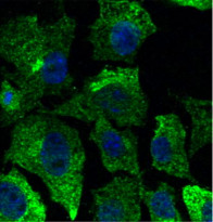
ARG42496 anti-Caveolin 1 antibody ICC/IF image
Immunofluorescence: Hepa1-6 cells were fixed with 4% PFA. Cells were stained with ARG42496 anti-Caveolin 1 antibody (green) at 1:50 dilution. Nuclear staining (blue).
-
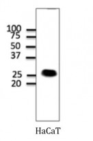
ARG42496 anti-Caveolin 1 antibody WB image
Western blot: 100 µg of HaCaT cell lysate stained with ARG42496 anti-Caveolin 1 antibody at 1:1000 dilution.


