ARG42684
anti-Cathepsin L + Cathepsin V antibody [33/2]
anti-Cathepsin L + Cathepsin V antibody [33/2] for IHC-Frozen sections,Western blot and Human,Mouse,Rat
Overview
| Product Description | Mouse Monoclonal antibody [33/2] recognizes Cathepsin L + Cathepsin V |
|---|---|
| Tested Reactivity | Hu, Ms, Rat |
| Tested Application | IHC-Fr, WB |
| Host | Mouse |
| Clonality | Monoclonal |
| Clone | 33/2 |
| Isotype | IgG1 |
| Target Name | Cathepsin L + Cathepsin V |
| Antigen Species | Human |
| Immunogen | Procathepsin L isolated from Human lung cancer cell line EPLC 32M1. |
| Conjugation | Un-conjugated |
| Alternate Names | Cathepsin L: Major excreted protein; CATL; Cathepsin L1; EC 3.4.22.15; CTSL1; MEP Cathepsin V: CTSU; Cathepsin L2; CTSL2; CATL2; Cathepsin U; EC 3.4.22.43 |
Application Instructions
| Application Suggestion |
|
||||||
|---|---|---|---|---|---|---|---|
| Application Note | * The dilutions indicate recommended starting dilutions and the optimal dilutions or concentrations should be determined by the scientist. |
Properties
| Form | Liquid |
|---|---|
| Purification | Unpurified |
| Buffer | Mouse ascites fluid, 1.2% Sodium acetate, 0.01% Sodium azide and 2% BSA. |
| Preservative | 0.01% Sodium azide |
| Stabilizer | 2% BSA |
| Concentration | 0.5 mg/ml |
| Storage Instruction | For continuous use, store undiluted antibody at 2-8°C for up to a week. For long-term storage, aliquot and store at -20°C or below. Storage in frost free freezers is not recommended. Avoid repeated freeze/thaw cycles. Suggest spin the vial prior to opening. The antibody solution should be gently mixed before use. |
| Note | For laboratory research only, not for drug, diagnostic or other use. |
Bioinformation
| Gene Symbol | CTSL; CTSV |
|---|---|
| Gene Full Name | cathepsin L; cathepsin V |
| Background | Cathepsin L: The protein encoded by this gene is a lysosomal cysteine proteinase that plays a major role in intracellular protein catabolism. Its substrates include collagen and elastin, as well as alpha-1 protease inhibitor, a major controlling element of neutrophil elastase activity. The encoded protein has been implicated in several pathologic processes, including myofibril necrosis in myopathies and in myocardial ischemia, and in the renal tubular response to proteinuria. This protein, which is a member of the peptidase C1 family, is a dimer composed of disulfide-linked heavy and light chains, both produced from a single protein precursor. Multiple alternatively spliced transcript variants have been found for this gene. [provided by RefSeq, Apr 2012] Cathepsin V: The protein encoded by this gene, a member of the peptidase C1 family, is a lysosomal cysteine proteinase that may play an important role in corneal physiology. This gene is expressed in colorectal and breast carcinomas but not in normal colon, mammary gland, or peritumoral tissues, suggesting a possible role for this gene in tumor processes. Alternatively spliced variants, encoding the same protein, have been identified. [provided by RefSeq, Jan 2011] |
| Function | Cathepsin L: Important for the overall degradation of proteins in lysosomes. [UniProt] Cathepsin V: Cysteine protease. May have an important role in corneal physiology. [UniProt] |
| Cellular Localization | Cathepsin L and Cathepsin V: Lysosome. [UniProt] |
| Calculated MW | Cathepsin L: 38 kDa Cathepsin V: 37 kDa |
Images (1) Click the Picture to Zoom In
-
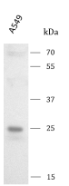
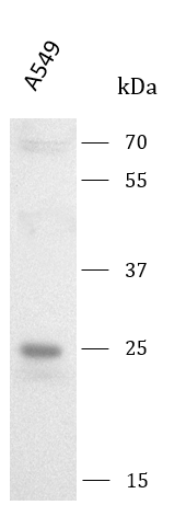
ARG42684 anti-Cathepsin L + Cathepsin V antibody [33/2] WB image
Western blot: A549 stained with ARG42684 anti-Cathepsin L + Cathepsin V antibody [33/2] at 1 μg/mL dilution.